L99-2 Review-Final
Lecture 7: Reproductive Behavior
Reproductive behaviors include:
- courting
- mating,
- parental behavior,
- most forms of aggressive behaviors
Coolidge effect:
- The restorative effect of introducing a new female sex partner to a male that has apparently become “exhausted” by sexual activity.
Hormonal Control of Sexual Behavior: Androgens, Testosterone, Estrogen(Oestrogen)
- Androgens(雄激素): are referred to as male sex hormones responsible for a number of male characteristics and functions.
- There are two main types of androgens namely, adrenal androgens(肾上腺雄激素) and testicular androgens(睾丸雄激素)
- Androgen activity is also present in females but in a very low scale. Androgens in females are involved in premature uterine contractions and help to create a balance of hormones.
- Testosterone(睾酮): the main androgen, which is the testicular androgen and is produced by the testes.
- testosterone is necessary for male sexual behavior
- the testosterone increases appear to be the result of sexual activity rather than the cause
- Estrogen(Oestrogen): are the main hormone that involves in imparting sexual characteristics to females. Estrogen is mainly secreted by the ovaries(卵巢). Similar to androgens, estrogen is also present in males but in very fewer quantities.
Brain Structures and Neurotransmitters
- Both sexes: the medial preoptic area (MPOA), the medial amygdala
- Stimulation of the MPOA increases copulation in rats of both sexes, and the MPOA is active when they copulate spontaneously.
- The MPOA appears to be more responsible for performance than for sexual motivation
- The amygdala is involved not only in sexual behavior but also in aggression and emotions. The medial amygdala is active while rats copulate, and stimulation causes the release of dopamine in the MPOA
- The medial amygdala’s role apparently is to respond to sexually exciting stimuli
- Stimulation of the MPOA increases copulation in rats of both sexes, and the MPOA is active when they copulate spontaneously.
- Males: sexually dimorphic nucleus (SDN)), paraventricular nucleus (PVN)
- SDN of the preoptic area (SDNPOA): Bigger in male. An characristic of male brain development. Associated with sexually dimorphic behaviors.
- The paraventricular nucleus (PVN,室旁核) is important for male sexual performance and, particularly, for penile erections.
- Female : ventromedial hypothalamus
- The ventromedial hypothalamus is active in females during copulation, and its destruction reduces the female’s responsiveness to a male’s advances.
- Role of oxytocin, testosterone, vasopressin
- Oxytocin:
- contributes to male and female orgasm and the intensity of its pleasure(性高潮以及快感强度)
- Intimacy increases oxytocin, but its interaction with testosterone levels determines whether that intimacy is sexual (if testosterone is high) or nurturing (if testosterone is low)
- Oxytocin determines the relative amount of social bonding versus social isolation
- Low testosterone with high oxytocin is correlated with more helpful, supporting intimacy and stronnger social bonds, where high levels of both hormones lead to more sexualized relationships with others.
- Therefore, the strongest social bonds result from high levels of oxytocin and vasopressin and low levels of testosterone.
- Testosterone:
- The effects of castration indicate that testosterone is necessary for male sexual behavior
- a high testosterone level in either sex increases aggression, but it also impairs the formation of close social bonds.
- Vasopressin:
- Antagonistic aggression(对抗性攻击) (which includes social dominance, partner acquisition, and defense of partners and territory) is seen in those with low levels of vasopressin.
- Protective aggression(保护性攻击) (such as defending children or partners) is seen in those with high levels of vasopressin.
- Oxytocin:
Pheromones:
- A chemical released by one animal that affects the behavior or physiology of another animal; usually smelled or tasted.
- Lee-Boot effect
- When groups of female mice are housed together, their estrous cycles slow down and eventually stop(发情期减慢并最终停止).
- The slowing and eventual cessation of estrous cycles in groups of female animals that are housed together; caused by a pheromone in the animals’ urine; first observed in mice.
- Whitten effect
- If groups of females are exposed to the odor of a male (or of his urine), they begin cycling again, and their cycles tend to be synchronized(开始发情周期且周期趋于同步)
- The synchronization of the menstrual or estrous cycles of a group of females, which occurs only in the presence of a pheromone in a male’s urine.
- Vandenbergh effect
- The Vandenbergh effect (Vandenbergh, Whitsett, and Lombardi, 1975) is the acceleration of the onset of puberty in a female rodent caused by the odor of a male.
- The earlier onset of puberty seen in female animals that are housed with males; caused by a pheromone in the male’s urine; first observed in mice.
- Bruce effect
- Termination of pregnancy caused by the odor of a pheromone in the urine of a male other than the one that impregnated the female; first identified in mice.
Difference between sex and gender
- Sex is the term for the biological characteristics that divide humans and other animals into the categories of male and female.
- Gender refers to the behavioral characteristics associated with being male or female.
Gender Identity
- gender identity is the person’s subjective feeling of being male or female.
Sexual orientation: biological basis
- Influenced by hormones: prenatal androgenization is responsible for this increased incidence of a masculinized sexual orientation
- Genes: Xq28
Parental Behavior
- Hormone: progesterone(黄体酮), estradiol(雌二醇 ), and prolactin(催乳素)
- Note that just before parturition the level of estradiol begins rising, then the level of progesterone falls dramatically, followed by a sharp increase in prolactin, the hormone produced by the anterior pituitary gland that is responsible for milk production
- The medial preoptic area (MPA), the region of the forebrain that plays the most critical role in male sexual behavior, appears to play a similar role in maternal behavior
- both prolactin and oxytocin facilitate a father’s role in infant care.
Lecture 8: Emotions
Discuss the behavioral, autonomic, and hormonal components of an emotional response and the role of the amygdala in controlling them.
- An emotional response consists of three types of components
- Behavioral component consists of muscular movements that are appropriate to the situation that elicits them.
- Autonomic responses facilitate the behaviors and provide quick mobilization of energy for vigorous movement.
- Hormonal responses reinforce the autonomic responses.
Negative emotions receive much more attention than positive ones: fear and anger
Amygdala is responsible to emotional responses (especially fear).
Fear
- Amygdala
- Central nucleus: Activation
- Lateral nucleus: Establishment of conditional emotional response
Conditioned Emotional Response: classically conditioned response that occurs when neutral stimulus is followed by an aversive stimulus(厌恶刺激).
Extinguish of conditioned emotional response:
- Lesions of the ventromedial prefrontal cortex (vmPFC) impair extinction
- Stimulation of this region inhibits conditioned emotional responses
Discuss the nature, functions, and neural control of aggressive behavior.
Anger, Aggression, and Impulse Control
Nature and functions of aggressive behavior:
- threatening gestures or actual attack directed toward another animal
- organized by neural circuits whose development is largely programmed by an animal’s genes
- related to reproduction
- related to self-defense
Neural Control of Aggressive Behavior:
- vmPFC plays a role in inhibiting emotional responses, such as aggression, violence, risk.
- activity of serotonergic synapses inhibits aggression.
- The neural control of aggressive behavior is hierarchical(分层的).
- The particular muscular movements an animal makes in attacking or defending itself are programmed by neural circuits in the brain stem.
- Whether an animal attacks depends on many factors, including the nature of the eliciting stimuli in the environment and the animal’s previous experience. The activity of the brain stem circuits appears to be controlled by the hypothalamus and the amygdala, which also influence many other species-typical behaviors.
- the activity of serotonergic synapses inhibits aggression.
Phineas Gage
- Prefrontal cortex plays a role in moral judgment.
- The amygdala plays an important role in provoking anger and violent emotional reactions, and the prefrontal cortex plays an important role in suppressing such behavior by making us see its negative consequences.
Discuss the role of the ventromedial prefrontal cortex in anger, aggression, and impulse control.
The ventromedial prefrontal cortex (vmPFC) plays a role in inhibiting emotional responses
- The vmPFC receives direct inputs from the dorsomedial thalamus, temporal cortex, ventral tegmental area, olfactory system, and amygdala.
- Its outputs go to several brain regions, including the cingulate cortex, hippocampal formation, temporal cortex, lateral hypothalamus, and amygdala.
- it communicates with other regions of the prefrontal cortex. Thus, its inputs provide it with information about what is happening in the environment and what plans are being made by the rest of the frontal lobes, and its outputs permit it to affect a variety of behaviors and physiological responses, including emotional responses organized by the amygdala.
Discuss cross-cultural studies on the expression and comprehension of emotions.
Cross-cultural studies:
- Facial expression of emotion is innate response
- The expression and comprehension of emotions are natural for our species and do not require learning by imitation
Discuss the neural control of the recognition of emotional expression.
We recognize other people’s feelings by means of vision and audition—seeing their facial expressions and hearing their tone of voice and choice of words.
- Laterality of Emotional Recognition:
- Right brain plays a more important role than the left hemisphere in comprehension of emotion.
- As for the emotion expression, the left half face (controlled by right brain) were more expressive than the right half face (controlled by left brain).
Amygdala:
- the amygdala plays a special role in emotional responses. It plays a role in emotional recognition as well
- Lesion of amygdala impair people’s ability to recognize facial expressions of emotion, especially expressions of fear
- amygdala lesions do not appear to affect people’s ability to recognize emotions in tone of voice.
- Amygdala is not involved in the expression of facial emotions.
Discuss the neural control of emotional expression.
The Mirror Neuron System:
- When we see a facial expression of an emotion, we unconsciously imagine ourselves making that expression. Often, we do more than imagine making the expressions—we actually imitate what we see.
- Mirror neurons are activated when an animal performs a particular behavior or when it sees another animal performing that behavior. These neurons are involved in learning to imitate the actions of others. These neurons, which are located in the ventral premotor cortex of the frontal lobe, receive input from the superior temporal sulcus and the posterior parietal cortex.
Communication of Emotions
- Amygdala is not involved in the expression of facial emotions.
- Role of imitation in recognition of emotion: The Mirror Neuron System
- If you can’t move your face, you are slower reading emotions
- Disease: Moebius syndrome, people affected with this syndrome cannot make facial expressions of emotion
- volitional facial paresis and emotional facial paresis
- volitional facial paresis is caused by damage to the face region of the primary motor cortex or to the fibers connecting this region with the motor nucleus of the facial nerve, which controls the muscles responsible for movement of the facial muscles.
- emotional facial paresis is caused by damage to the insular region of the prefrontal cortex, to the white matter of the frontal lobe, or to parts of the thalamus. This system joins the system responsible for voluntary movements of the facial muscles in the medulla or caudal pons.
Discuss the James-Lange theory of feelings of emotion and evaluate relevant research.
Feelings of emotion: James-Lange theory
- Behaviors and physiological responses are directly elicited by situations;
- Feelings of emotions are produced by feedback from these behaviors and responses.
Own emotional feelings are based on what we find ourselves doing and on sensory feedback we receive from the activity of our muscles and internal organs.
Other researches:
- Feedback from Simulated Emotions
- Several experiments suggest that feedback from the contraction of facial muscles can affect people’s moods and even alter the activity of the autonomic nervous system.
- Perhaps the connection is a result of experience; in other words, perhaps the occurrence of particular facial movements along with changes in the autonomic nervous system leads to classical conditioning, so that feedback from the facial movements becomes capable of eliciting the autonomic response—and a change in perceived emotion.
Lecture 9: Ingestive Behavior
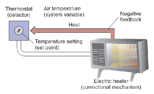
Explain the characteristics of a regulatory mechanism.
A regulatory mechanism contains four essential features:
- the system variable (the characteristic to be regulated),
- a set point (the optimal value of the system variable),
- a detector that monitors the value of the system variable,
- a correctional mechanism that restores the system variable to the set point
- Negative feedback is a process whereby the effect produced by an action serves to diminish or terminate that action; a characteristic of regulatory systems.
Describe the fluid compartments of the body.
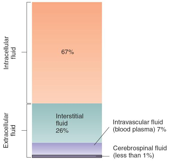
Two of the body’s fluid compartments must be kept within precise limits: the intracellular fluid and the intravascular fluid
- The intracellular fluid is controlled by the concentration of solutes in the interstitial fluid.
Explain the control of osmometric thirst and volumetric thirst and the role of angiotensin.
Loss of water from either the intracellular or intravascular fluid compartments stimulates drinking, researchers have adopted the terms osmometric thirst (渗透性口渴) and volumetric thirst(体积性口渴) to describe them.
Thirst produced by an increase in the osmotic pressure of the interstitial fluid relative to the intracellular fluid, thus producing cellular dehydration (the cells, and they shrink in volume).
- The detector is responding to (metering) changes in the concentration of the interstitial fluid that surrounds them.
- Actin filaments in brain osmoreceptors detect changes in solute concentration in the interstitial fluid when the cell membrane expands or contracts. Changes in cell volume cause changes in the membrane potential, which serve as the signal for osmometric thirst.
- Most researchers now believe that osmoreceptors responsible for osmometric thirst are located in the region of the anterior hypothalamus that borders the anteroventral tip of the third ventricle (the AV3V).
Volumetric Thirst:
- Volumetric thirst occurs when the volume of the blood plasma (the intravascular volume) decreases.
- evaporation produces both volumetric thirst and osmometric thirst.
- In addition, loss of blood, vomiting, and diarrhea all cause loss of blood volume (hypovolemia) without depleting the intracellular fluid.
- Volumetric thirst is produced by hypovolemia.
Role of Angiotensin 血管紧张素
- A peptide hormone that constricts blood vessels, causes the retention of sodium and water, and produces thirst and a salt appetite.
- The kidneys contain cells that are able to detect decreases in the flow of blood to the kidneys.
- The usual cause of a reduced flow of blood is a loss of blood volume; thus, these cells detect the presence of hypovolemia.
- When the flow of blood to the kidneys decreases, these cells secrete an enzyme called renin(肾素).
- Renin enters the blood, where it catalyzes the conversion of a protein called angiotensinogen into a hormone called angiotensin(血管紧张素).
- Angiotensin II has several physiological effects:
- It stimulates the secretion of hormones by the posterior pituitary gland and the adrenal cortex that cause the kidneys to conserve water and sodium;
- It increases blood pressure by causing muscles in the small arteries to contract.
- Angiotensin II has two behavioral effects:
- It initiates both drinking and a salt appetite. Therefore, a reduction in the flow of blood to the kidneys causes water and sodium to be retained by the body, helps to compensate for their loss by reducing the size of the blood vessels, and encourages the animal to find and ingest both water and salt
The kidneys contain cells that are able to detect decreases in the flow of blood to the kidneys
- Renin: A hormone secreted by the kidneys that causes the conversion of angiotensinogen in the blood into angiotensin.
- Angiotensin: A peptide hormone that constricts blood vessels, causes the retention of sodium and water, and produces thirst and a salt appetite.
- Subfornical organ (SFO): detect the presence of angiotensin
- Median preoptic nucleus : plays a role in thirst stimulated by angiotensin.
Describe characteristics of the two nutrient reservoirs and the absorptive and fasting phases of metabolism.
There are two reservoirs: one short-term and the other long-term. The short-term reservoir stores carbohydrates, and the long-term reservoir stores fats.
- The short-term reservoir is located in the cells of the liver and the muscles. For simplicity we will consider only one of these locations: the liver. Cells in the liver convert glucose (a simple, soluble carbohydrate) into glycogen and store the glycogen. They are stimulated to do so by the presence of insulin , a peptide hormone produced by the pancreas.
- Thus, when glucose and insulin are present in the blood, some of the glucose is used as a fuel, and some of it is stored as glycogen. Later, when all of the food has been absorbed from the digestive tract, the level of glucose in the blood begins to fall.
- The fall in glucose is detected by cells in the pancreas and in the brain. The pancreas responds by stopping its secretion of insulin and starting to secrete a different peptide hormone: glucagon . The effect of glucagon is opposite that of insulin: It stimulates the conversion of glycogen into glucose.
- Thus, the liver soaks up excess glucose and stores it as glycogen when plenty of glucose is available, and it releases glucose from its reservoir when the digestive tract becomes empty and the level of glucose in the blood begins to fall.
- Our long-term reservoir consists of adipose tissue (fat tissue). This reservoir is filled with fats, or, more precisely, with triglycerides . Adipose tissue is found beneath the skin and in various locations in the abdominal cavity. It consists of cells that are capable of absorbing nutrients from the blood, converting them to triglycerides, and storing them. These cells can expand enormously in size
- when we wake in the morning with an empty digestive tract, our brain is living on glucose released by the liver. But what about the other cells of the body? They are living on fatty acids, sparing the glucose for the brain.
- When the digestive system is empty, there is an increase in the activity of the sympathetic axons that innervate adipose tissue, the pancreas, and the adrenal medulla. All three effects (direct neural stimulation, secretion of glucagon, and secretion of catecholamines) cause triglycerides in the long-term fat reservoir to be broken down into glycerol and fatty acids.
Fasting phase
- The phase of metabolism during which nutrients are not available from the digestive system; glucose, amino acids, and fatty acids are derived from glycogen, protein, and adipose tissue during this phase.
Absorptive phase
- The phase of metabolism during which nutrients are absorbed from the digestive system; glucose and amino acids constitute the principal source of energy for cells during this phase, and excess nutrients are stored in adipose tissue in the form of triglycerides.
Eating
- The short-term reservoir stores carbohydrates in the liver, and the long-term reservoir stores fats.
- Cells in the liver convert glucose into glycogen and store the glycogen. They are stimulated to do so by the presence of insulin , a peptide hormone produced by the pancreas.
- The glucose is primarily transported to the brain as the fuel, because of insulin: their glucose transporters do not contain insulin receptors.
Discuss the signals from the environment, the stomach, and the metabolism that begin a meal.
Signals from the Stomach:
- An empty stomach and upper intestine provide an important signal to the brain that it is time to start thinking about finding something to eat.
- The gastrointestinal system (especially the stomach) releases a peptide hormone called ghrelin (生长激素释放激素、饥饿激素)
- The blood levels of ghrelin increase with fasting and are reduced after a meal.
- Although ghrelin is an important short-term hunger signal, it clearly cannot be the only one.
Signals from the Environment:
- Although an empty stomach is an important signal, many factors start a meal, including the sight of a plate of food, the smell of food cooking in the kitchen, the presence of other people sitting around the table, or the words “It’s time to eat!”
Metabolic signals:
- A fall in blood glucose level (a condition known as hypoglycemia) is a potent stimulus for hunger.
- Hunger can also be produced by causing lipoprivation —depriving cells of lipids. More precisely, they are deprived of the ability to metabolize fatty acids through injection of a drug that interferes with this process.
Discuss the long-term and short-term factors that stop a meal.
Short-term satiety signals come from the immediate effects of eating a particular meal, which begin long before the food is digested.
Long-term satiety signals arise in the adipose tissue, which contains the long-term nutrient reservoir.
Summary for starting a meal and stopping a meal
What starts a meal and what stops a meal?
- Ghrelin:A peptide hormone released by gastrointestinal system (especially the stomach) that initiate eating.
- Cholecystokinin (CCK): A peptide hormone released by duodenum provide a satiety signal to the brain.
- CCK does not act directly on the brain; instead, it acts on receptors located in the junction between the stomach and the duodenum
- PYY:secreted by cells in the gastrointestinal tract that serves as an additional satiety signal.
- Only nutrients caused PYY to be secreted; a large drink of water had no effect
- Insulin: secreted by pancreas and serve as a satiety signal.
- Insulin permits organs other than the brain to metabolize glucose.
- Insulin promotes the entry of nutrients into fat cells where they are converted into triglycerides.
- Insulin is a peptide and would not normally be admitted to the brain. However, a transport mechanism delivers it through the blood–brain barrier, and it reaches neurons in the hypothalamus that are involved in regulation of hunger and satiety.
- Leptin: secreted by well-nourished fat cells, acting as an antiobesity hormone.
- Long-term satiety.
- A particular gene, called OB, normally produces a peptide hormone that has been given the name leptin(瘦素)
- If ob mice are given daily injections of leptin, their metabolic rate increases, their body temperature rises, they become more active, and they eat less. As a result, their weight returns to normal.
| Name | Where Produced | Site of Actions | Physiological or Behavioral Effects |
|---|---|---|---|
| Leptin | Fat tissue | Inhibits NPY/AGRP neurons; excites CART/α-MSH neurons | Suppression of eating, increased metabolic rate |
| Insulin | Pancreas | Similar to leptin | Similar to leptin |
| Ghrelin | Gastrointestinal system | Activates NPY/AGRP neurons | Eating |
| Cholecystokinin (CCK) | Duodenum | Neurons in pylorus | Suppression of eating |
| Peptide YY3–36 (PYY) | Gastrointestinal system | Inhibits NPY/AGRP neurons | Suppression of eating |
Describe research on the role of the brain stem and hypothalamus in hunger and satiety.
The brain stem contains neural circuits that can detect hunger and satiety signals and control at least some aspects of food intake.
- wo regions of the medulla, the area postrema and the nucleus of the solitary tract (henceforth referred to as the AP/NST), receive taste information from the tongue and a variety of sensory information from the internal organs, including signals from detectors in the stomach, duodenum, and liver.
- In addition, this region contains a set of detectors that are sensitive to the brain’s own fuel: glucose. All this information is transmitted to regions of the forebrain that are more directly involved in control of eating and metabolism.
- Evidence indicates that events that produce hunger increase the activity of neurons in the AP/NST. In addition, lesions of this region abolish both glucoprivic and lipoprivic feeding.
two regions of the hypothalamus: the lateral area and the ventromedial area.
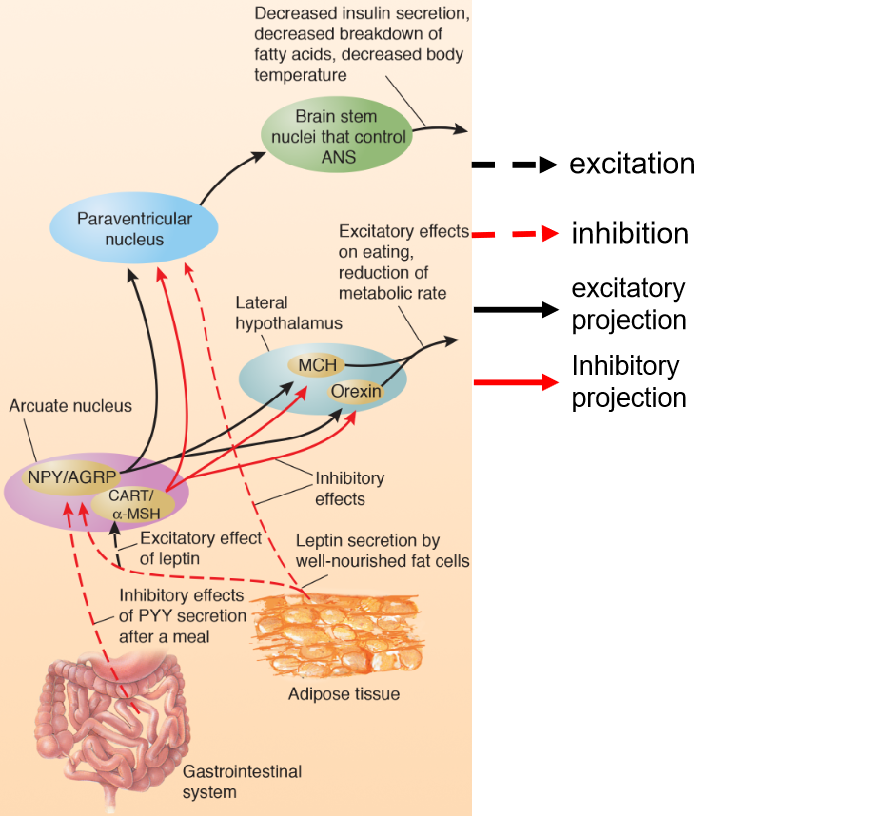
| Name | Location of Cell Bodies | Location of Terminals | Interaction with Other Peptides | Physiological or Behavioral Effects |
|---|---|---|---|---|
| Melanin-concentratinghormone (MCH) | Lateral hypothalamus | Neocortex, periaqueductal gray matter, reticular formation, thalamus, locus coeruleus, neurons in spinal cord that control the sympathetic nervous system | Activated by NPY/AGRP; inhibited by leptin and CART/α-MSH | Eating, decreased metabolic rate |
| Orexin | Lateral hypothalamus | Similar to those of MCH neurons | Activated by NPY/AGRP;inhibited by leptin and CART/α-MSH | Eating, decreased metabolic rate |
| Neuropeptide Y (NPY) | Arcuate nucleus of hypothalamus | Paraventricular nucleus, MCH and orexin neurons of the lateral hypothalamus | Activated by ghrelin;inhibited by leptin | Eating, decreased metabolic rate |
| Agouti-related protein (AGRP) | Arcuate nucleus of hypothalamus (colocalized with NPY) | Same regions as NPY neurons | Inhibited by leptin | Eating, decreased metabolic rate; acts as antagonist atMC4 receptors |
| Cocaine- and amphetamine-regulated transcript(CART) | Arcuate nucleus of hypothalamus | Paraventricular nucleus, lateral hypothalamus, periaqueductal gray matter, neurons in spinalcord that control the sympathetic nervous system | Activated by leptin | Suppression of eating, increased metabolic rate |
| α-Melanocyte stimulating hormone (α-MSH) | Arcuate nucleus of hypothalamus(colocalized withCART) | Same regions as CART neurons | Activated by leptin | Suppression of eating, increased metabolic rate; actsas agonist at MC4 receptors |
Discuss the social and physiological factors that contribute to obesity.
Genetic differences—and their effects on development of the endocrine system and brain mechanisms that control food intake and metabolism—appear to be responsible for the overwhelming majority of cases of extreme obesity.
people’s behavior (availability of cheap, tasty, calorie-rich food and decreased levels of exercise).
Discuss surgical, behavioral, and pharmacological treatments for obesity.
Bariatric surgery are designed to reduce the amount of food that can be eaten during a meal or interfere with absorption of calories from the intestines. Bariatric surgery has been aimed at the stomach, the small intestine, or both.
- Gastric Sleeve, Gastric Bypass and LAP-BAND®
There are three possible ways in which drugs could help people lose weight:
- reduce the amount of food they eat
- prevent some of the food they eat from being digested
- increase their metabolic rate (that is, provide them with a “spendthrift phenotype”).
Eating disorder: Obesity
- Causes: environmental causes, hereditary factors, an efficient metabolism, inefficient metabolism
- Treatment: Bariatric surgery, exercise, drug treatment (not good)
Discuss the physiological factors that may contribute to anorexia nervosa and bulimia nervosa.
Anorexia nervosa and Bulimia nervosa
- Anorexics express an intense fear of becoming obese, but not lose interest in food.
The obsessions with food and weight loss and the compulsive rituals that people with anorexia nervosa develop suggest a possible linkage with obsessive-compulsive disorder.
- Many anorexics exercise by cycling, running, or almost constant walking and pacing.
Treatment:
- Cognitive behavior therapy
Food is a natural reinforcer.
Reward refers to the positive effect an object or a condition—such as a drug, food, sexual contact, or warmth—has on the user.
Reward circuit: the mesolimbic pathway, dopaminergic neurons originating in the ventral tegmental area and connecting to several targets in the limbic system, especially the nucleus accumbens.
Drug Abuse
- Addictive drugs “hijack” the reward brain mechanisms, produces long-term changes in the brain.
Drug tolerance: decreased sensitivity to a drug.
Withdraw symptoms : primarily the opposite of the effects of the drug itself.
Related brain region: prefrontal cortex.
- Stressful situations can cause former drug addicts to relapse.
- The stress-induced release of corticotropin-releasing hormone (CRH) caused an enhanced activation of dopaminergic neurons in the ventral tegmental area in mice that had been exposed to cocaine.
Causes: both genetic and environmental factors
| Drug | Sites of Action |
|---|---|
| Ethyl alcohol | NMDA receptor (indirect antagonist); GABAA receptor(indirect agonist) |
| Barbiturates | GABAA receptor (indirect agonist) |
| Benzodiazepines (tranquilizers) | GABAA receptor (indirect agonist) |
| Cannabis (marijuana) | CB1 cannabinoid receptor (agonist) |
| Nicotine | Nicotinic ACh receptor (agonist) |
| Opiates (heroin, morphine, etc.) | μ and δ opiate receptor agonist |
| Phencyclidine (PCP) and ketamine | NMDA receptor (indirect antagonist) |
| Cocaine | Blocks reuptake of dopamine (and serotonin andnorepinephrine) |
| Amphetamine | Causes release of dopamine (by running dopaminetransporters in reverse) |
Lecture 10: Learning and Memory
Describe each of four basic forms of learning: perceptual learning, stimulus–response learning, motor learning, and relational learning.
- perceptual learning 知觉学习
- Learning to recognize a particular stimulus. 学习识别特定的刺激
- stimulus–response learning 刺激反应学习
- Learning to automatically make a particular response in the presence of a particular stimulus; includes classical and instrumental conditioning. 学习在特定反应条件下做出特定的刺激
- motor learning 运动学习
- Learning to make a new response. 学习做出新的反应
- Motor learning: Learning to make a new response. Establishment of changes within motor systems
- Motor learning, is actually a component of stimulus– response learning.
- relational learning 关联学习
- Learning the relationships among individual stimuli. 学习单个刺激之间的关系
- Episodic learning, hippocampus
a defensive eyeblink response can be conditioned to a tone. If we direct a brief puff of air toward a rabbit’s eye, the eye will automatically blink. The response is called an unconditional response (UR) because it occurs unconditionally, without any special training. The stimulus that produces it (the puff of air) is called an unconditional stimulus (US)
Classical conditioning is a form of learning in which an unimportant stimulus acquires the properties of an important one.. Classical conditioning has occurred; the conditional stimulus (CS —the 1000-Hz tone) now elicits the conditional response (CR–the eye blink).
Instrumental (operant) conditioning : adjust the behavior according to the consequences.
- Instrumental conditioning is also called operant conditioning.
- Instrumental conditioning is a more flexible form of learning. It permits an organism to adjust its behavior according to the consequences of that behavior.
Hebb’s rule: fire together, wire together
- if a synapse repeatedly becomes active at about the same time that the postsynaptic neuron fires, changes will take place in the structure or chemistry of the synapse that will strengthen it.
Reinforcing stimulus, punishing stimulus, positive/negative reinforcement, positive/negative punishment
- Collectively, “favorable consequences” are referred to as reinforcing stimuli , and “unfavorable consequences” are referred to as punishing stimuli .
Describe the anatomy of the hippocampus, describe the establishment of long-term potentiation, and discuss the role of NMDA receptors in this phenomenon.
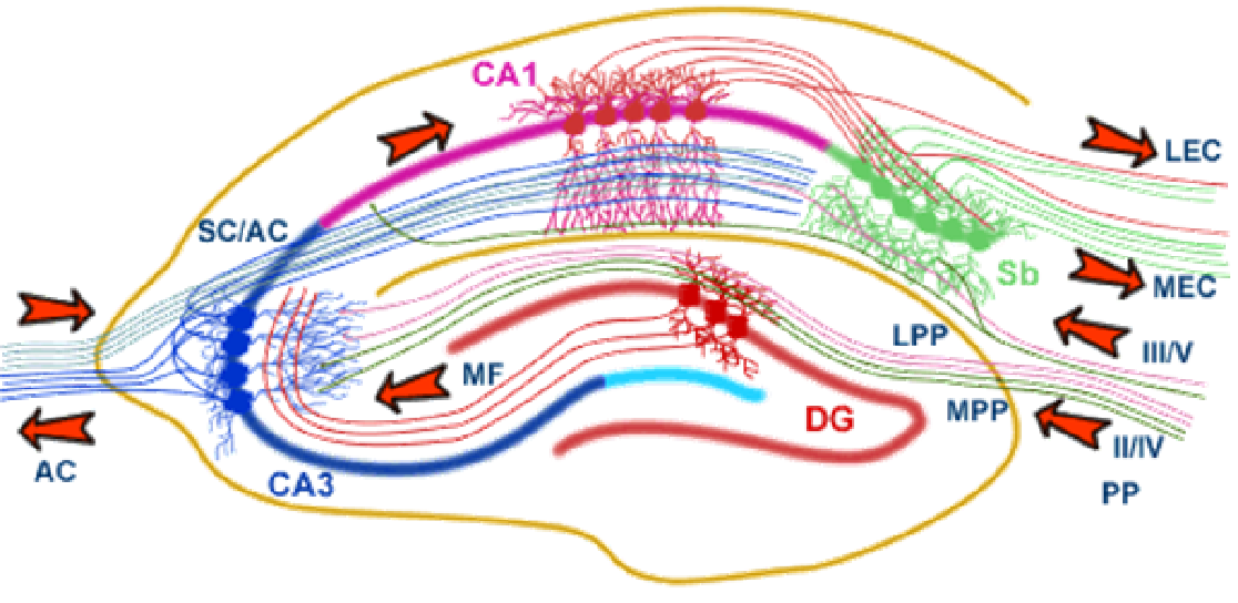
The anatomy and circuitry of the Hippocampus:
- entorhinal cortex → dentate gyrus →CA3 → CA1
long-term potentiation:
- A long-term increase in the excitability of a neuron to a particular synaptic input caused by repeated high-frequency activity of that input.
Long-term potentiation can be produced in other regions of the hippocampal formation and in many other places in the brain.
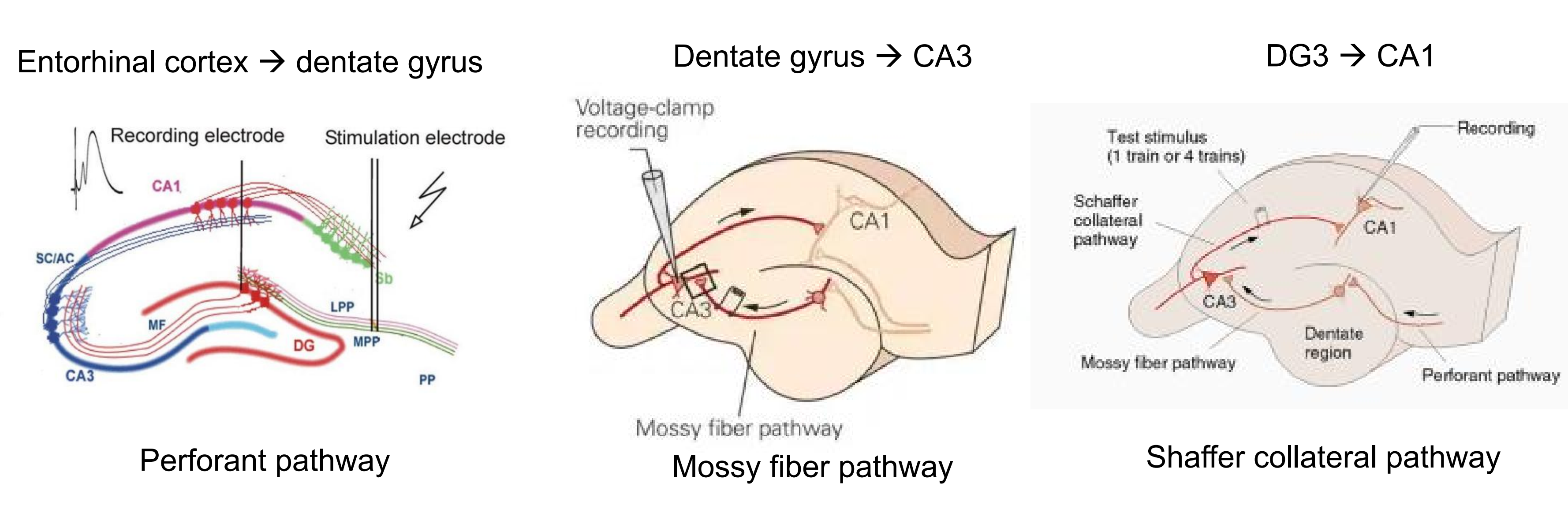
- A long-term potentiation in which concurrent stimulation of weak and strong synapses to a given neuron strengthens the weak ones.
Long-term potentiation requires two events: activation of synapses and depolarization of the postsynaptic neuron.
- The explanation for this phenomenon lies in the characteristics of a very special type of glutamate receptor: the NMDA receptor (N-methyl-D-aspartate).
- The NMDA receptor controls a calcium ion channel.
Discuss research on the physiological basis of synaptic plasticity during long-term potentiation and long-term depression.
Synaptic plasticity
- Synaptic plasticity: long-term potentiation (LTP) and long- term depression (LTD).
Long-term potentiation :
- Two types of LTP
- Associative LTP: when weak and strong synapses to a single neuron are stimulated at approximately the same time, the weak synapse becomes strengthened (Hebb’s Rule).
- Homosynaptic LTP: a high rate all in one burst will produce long-term potentiation of the synapse strength.
- Activation of synapses (presynaptic glutamate release) & depolarization of the postsynaptic neuron.
- NMDA receptor is calcium permeable
- NMDA receptor is transmitter (glutamate) and voltage dependent.
- AMPA receptors move to the synapses.
- Early phase LTP (E-LTP) and late phase LTP (L-LTP)
- Late phase LTP need protein synthesis
- PKM-zeta is an important enzyme involved in L-LTP
- A positive feedback.
- The self-sustaining synthesis of PKM-zeta makes long-term LTP possible.
Long-term depression:
- Low-frequency stimulation of the synaptic inputs to a cell can decrease their synaptic strength.
- LTD appears to involve the opposite: a decrease in the number of AMPA receptors in these spines.
Describe research on the role of the inferior temporal cortex in visual perceptual learning.
people with damage to the inferior temporal cortex may have excellent vision but be unable to recognize familiar, everyday objects such as scissors, clothespins, or light bulbs—as well as faces of friends and relatives.
- The ventral stream, which is involved with object recognition, continues ventrally into the inferior temporal cortex.
Discuss the physiology of the classically conditioned emotional response to aversive stimuli.
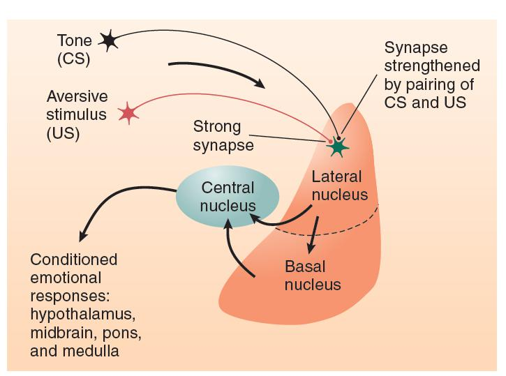
lateral nucleus → basal nucleus → central nucleus → conditioned emotional responses, hypothalamus, midbrain, pons, medulla
Describe the role of the basal ganglia in instrumental conditioning.
Instrumental conditioning entails the strengthening of connections between neural circuits that detect a particular stimulus and neural circuits that produce a particular response.
There are two major pathways between the sensory association cortex and the motor association cortex:
- direct transcortical connections (connections from one area of the cerebral cortex to another)
- connections via the basal ganglia and thalamus.
Studies with laboratory animals have found that lesions of the basal ganglia disrupt instrumental conditioning but do not affect other forms of learning.
Describe the role of dopamine in reinforcing brain stimulation and discuss the effects of administering dopamine antagonists and agonists.
Activity of dopaminergic neurons plays a particularly important role in reinforcement.
The mesolimbic system of dopaminergic neurons begins in the ventral tegmental area (VTA) of the midbrain and projects rostrally to several forebrain regions, including the amygdala, hippocampus, and nucleus accumbens (NAC) .
Neurons in the NAC project to the ventral part of the basal ganglia, which, as we just saw, are involved in learning.
The mesocortical system also plays a role in reinforcement. In addition, this system begins in the ventral tegmental area but projects to the prefrontal cortex, the limbic cortex, and the hippocampus.
Describe the nature of human anterograde amnesia and explain what it suggests about the organization of learning.
The term anterograde amnesia refers to difficulty in learning new information. A person with pure anterograde amnesia can remember events that occurred in the past, from the time before the brain damage occurred, but cannot retain information about events encountered after the damage.
confabulation may be caused by brain damage that disrupts the normal functions of the prefrontal cortex.
The surgery successfully treated H. M.’s seizure disorder, but it became apparent that the operation had produced a serious memory impairment. Further investigation revealed that the critical site of damage was the hippocampus.
Milner and her colleagues based the following conclusions on H.M’s pattern of deficits:
- The hippocampus is not the location of long-term memories; nor is it necessary for the retrieval of long-term memories.
- The hippocampus is not the location of immediate (short-term) memories.
- The hippocampus is involved in converting immediate (short-term) memories into long-term memories.
Describe the role of the hippocampus in relational learning, including episodic and spatial learning, and discuss the function of hippocampal place cells.
Without the hippocampal formation we would be left with individual, isolated memories without the linkage that makes it possible to remember—and think about—episodes and contexts.
Something about H.M.:
- H.M was still able to remember episodic memories before surgery.
- H.M was capable of other forms of learning: perceptual learning, stimulus-resp learning and motor learning.
- H.M could not form spatial memory
- Place cells in the hippocampus
- Hippocampus as a cognitive map
- Morris Water Maze: relational task (variable start position) and stimulus-response task (constant start position)
Episodic learning—remembering sequences of events (episodes) that we witness—requires us to keep track of and remember not only individual events but also the order in which they occur.
- A special system that involves the hippocampus and associated structures appears to perform coordinating functions required for many types of learning that go beyond simple perceptual, stimulus–response, or motor learning.
Lecture 11: Psychological Disorders
Schizophrenia 精神分裂症
- Definition: break with reality caused by disorganization of the various functions of the mind, such that thoughts and feelings no longer worked together normally.
- It does not mean multiple personality.
- Symptoms:
- Positive: delusions(幻想,错觉), hallucinations(幻觉), or thought disorders.
- Negative: flattened emotional response, poverty of speech, lack of initiative and persistence, anhedonia(快感缺乏), and social withdrawal.
- Cognitive: sustaining attention, low psychomotor speed, deficits in learning and memory, poor abstract thinking, and poor problem solving.
- Causes: Heritability (DISC1)
- Positive symptoms:
- The dopamine hypothesis suggests that the positive symptoms of schizophrenia are caused by over-activity of DA synapses.
- Related brain regions: mesolimbic pathway (tegmental area, the nucleus accumbens(伏隔核), amygdala)
- Treatment : chlorpromazine (氯丙嗪,dopamine antagonist)
- Negative and cognitive symptoms:
- Related brain region: brain abnormalities in the prefrontal cortex
- Treatment:
- atypical antipsychotic medications(非典型性精神病药), such as Clozapine(氯氮平)
- Third generation” antipsychotic drug, (e.g.aripiprazole(阿立哌唑)), partial agonist
- Schizophrenia is a developmental disease, but not caused by a degenerative process
Major Affective Disorders 情感障碍
- Major Depressive Disorder (MDD): characterized by depression without mania(躁狂). This depression may be continuous and unremitting or, more typically, may come in episodes.
- Biological Treatments: drugs that inhibit the reuptake of norepinephrine or serotonin, electroconvulsive therapy (ECT)transcranial magnetic stimulation (TMS), deep brain stimulation (DBS), bright-light therapy (phototherapy), sleep deprivation.
- Related brain region: Frontal cortex, amygdala, hippocampus
- Role of Circadian Rhythms in depression.
- One of the most prominent symptoms of depression is disordered sleep.
- Bipolar disorder: characterized by alternating periods of mania and depression.
- Biological Treatments for bipolar disorder: Lithium
Anxiety Disorders
- Types: Panic attacks (anticipatory anxiety, agoraphobia), generalized anxiety disorder, Social anxiety disorder
- Causes : hereditary component, BDNF (brain-derived neurotrophic factor)
- Related brain regions: amygdala and the cingulate, prefrontal, and insular cortices.
- Treatment: benzodiazepines (agonists of GABAA receptors ), serotonin agonists (e.g. fluoxetine)
Obsessive-Compulsive Disorder
- Symptoms: People with an obsessive-compulsive disorder (OCD) suffer from obsessions : (thoughts that will not leave them), and compulsions: behaviors that they cannot keep from performing.
- Causes:
- Heritability
- brain damage:basal ganglia, cingulate gyrus, and prefrontal cortex
- Treatment:
- Cingulotomy: The surgical destruction of the cingulum bundle, which connects the prefrontal cortex with the limbic system; helps to reduce intense anxiety and the symptoms of obsessive-compulsive disorder
- fluoxetine (serotonergic agonists)
Autistic Disorder
- Description: What is the symptoms of autistic disorder?
- Failure to develop normal social relations with other people,
- Impaired development of communicative ability,
- Repetitive, stereotyped movements.
- Most people with autistic disorder display cognitive impairments.
- Possible Causes: biological origin (Heritability, Brain Pathology )
- Related brain area: frontal cortex, temporal cortex, fusiform face area (FFA)
- Oxytocin
Attention-Deficit/Hyperactivity Disorder
- Symptoms : A disorder characterized by uninhibited responses, lack of sustained attention, and hyperactivity
- Related brain regions: prefrontal cortex
- Treatment: dopamine agonist (methylphenidate)
Stress Disorders
- Physiological definition of the stress: the physiological reaction caused by the perception of aversive or threatening situations.
- Stress hormone:
- Epinephrine: affects glucose metabolism, causing the nutrients stored in muscles to become available to provide energy
- Norepinephrine: increases blood flow
- Glucocorticoid: down protein and convert it to glucose, help to make fats available for energy, increase blood flow, and stimulate behavioral responsiveness, suppresses the secretion of the sex steroid hormones…
- Corticotropin-releasing hormone(CRH) → adrenocorticotropic hormone (ACTH) → Glucocorticoid
- Health Effects of Long-Term Stress
- Caused by prolonged secretion of glucocorticoids
- Increased blood pressure, damage to muscle tissue, steroid diabetes, infertility, inhibition of growth, inhibition of the inflammatory responses, and suppression of the immune system.
- Effects of Stress on the Brain: brain damage (hippocampus, amygdala, prefrontal cortex)
- Posttraumatic Stress Disorder 创伤后应激障碍
- Related gene:dopamine D2 receptors, dopamine transporters, and 5-HT transporters.
- Related brain region:amygdala, prefrontal cortex.
- Treatment: cognitive behavior therapy, group therapy, and SNRIs.