L2 Nervous System Development and Diseases
一、Early nervous system development
Formation of nervous system
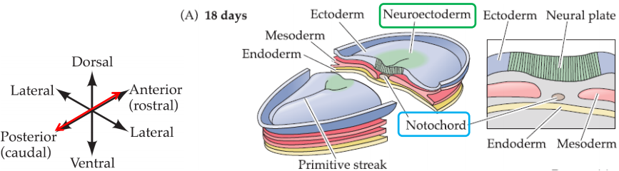
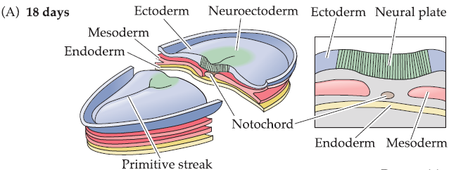
1. Gastrulation 原肠胚形成
Local invagination: three germ layers
- Outer ectoderm (外胚层,外细胞层)
- Middle mesoderm (中胚叶;中胚层) (initiating invagination (入鞘;套入))
- Inner endoderm (内胚层)
Establishment of the midline and the basic body axes
Formation of notochord (脊索) at the midline: central event
- Notochord: distinct cylinder of mesodermal cells
- Notochord defines embryonic midline and is the axis of symmetry for the entire body
- Neuroectoderm (神经外胚层 ): lies immediately above the notochord and gives rise to the entire nervous system
- Notochord: transient structure that disappears once early development is complete
2. Neurulation 神经胚形成
Notochord sends inductive signals to the overlaying ectoderm that cause a subset of cells to differentiate into neuroectodermal precursor cells.
Midline ectoderm containing these cells thickens into a distinct columnar (柱形的,筒形的) epithelium called neural plate (神经板).
The lateral margins of the neural plate fold inward, transforming the neural plate into a tube—neural tube (神经管).
The cells in neural tube gives rise to the brain, the spinal cord and most of PNS.
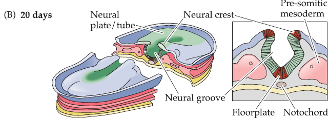
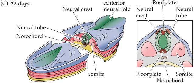
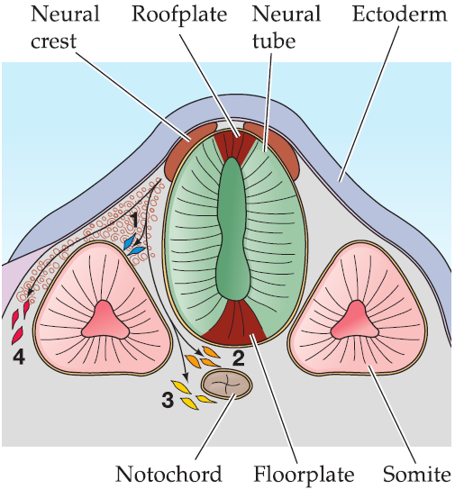
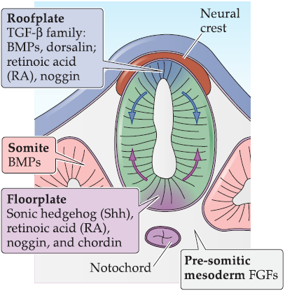
Floor plate (底板) and roof plate (顶板)
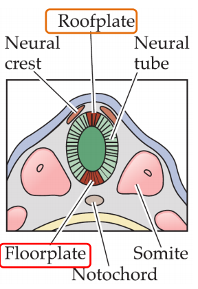
(1) Floor Plate 底板
Floor plate: specialized strip of epithelial-like cells at the ventral midline of the neural tube. Sequential functions as follows:
Molecular signals from the floor plate as well as from the notochord specify position and fate for the neuroectodermal precursors of the spinal cord and hindbrain.
These signals lead to differentiation of cells in the ventral (腹的,腹部的,腹侧的) portion of the neural tube that eventually give rise to spinal and hindbrain motor neurons and related interneurons (details later on)
(2) Roof Plate 顶板
Roof plate: narrow strip of neuroepithelial cells at the dorsal (背的,背侧的;背部的) midline of the neural tube.
- Molecular signals from the roof plate help define position and fate for the interneurons in the more dorsal regions of the spinal cord and hindbrain (details later on).
Neural crest 神经脊
神经脊(英语:neural crest)属于动物胚胎的外胚层,位于神经管(neural tube)与胚胎上皮之间。神经脊细胞在神经板形成(neurulation)之后不久就会离开原来所在位置。
Neural crest: the region where the edges of the folded neural plate come together.
Neural crest cells: a third population of precursor cells.
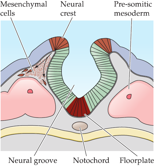
Migrating away from the neural tube through a matrix of loosely packed mesenchymal cells (间充质细胞).
Subsets of neural crest cells follow different pathways, along which they are exposed to additional signals that influence their specific differentiation, and give rise to:
- Neurons and glia of the sensory ganglia.
- Neurons and glia of the visceral motor (autonomic) ganglia.
- Neurosecretory cells of the adrenal gland which eventually aggregate around the dorsal portion of the kidney.
- Non-neural structures such as pigment cells, cartilage, and bone, particularly in the face and skull.
从Neuro crest来的细胞经过不同的组织,接受这些组织释放信号,从而决定这些从Neuro crest细胞的分化情况
Neural stem cells
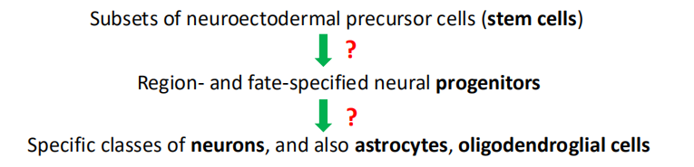
1. Neuroectodermal precursor cells: neural stem cells
神经外胚层前体细胞
Dividing to produce more precursor cells (self-renewal)
Capable to give rise to the full range of cell classes
2. Differentiation order
- Subsets of neuroectodermal precursor cells (stem cells)
- Region- and fate-specified neural progenitors (先驱)
- Specific classes of neurons, and also astrocytes, oligodendroglial cells
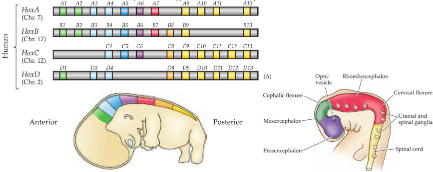
二、Formation of Major Brain Subdivisions
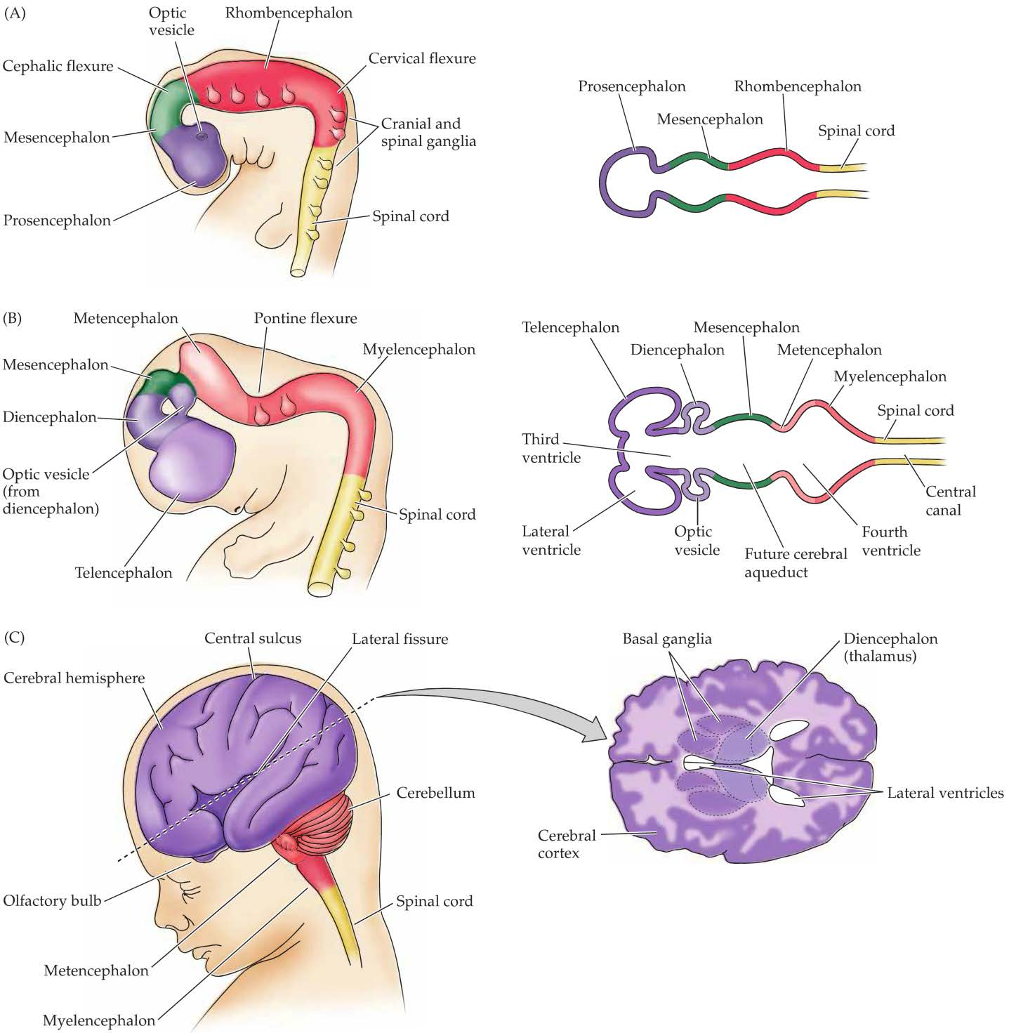
Segmentation 分割
The neural tube is organized into repeating units called neuromeres (神经管节).
Segmentation: establishing regional identity in the body by dividing the embryo into repeated units, or segments.
Fruit fly Drosophila: early expression of homeotic or homeobox genes guides the differentiation of the embryo into distinct segments that give rise to the head, thorax (胸,胸膛;胸腔), and abdomen (腹部;下腹;腹腔).

- Temporal (短暂的) pattern of expression of four homeobox genes encoding TFs.
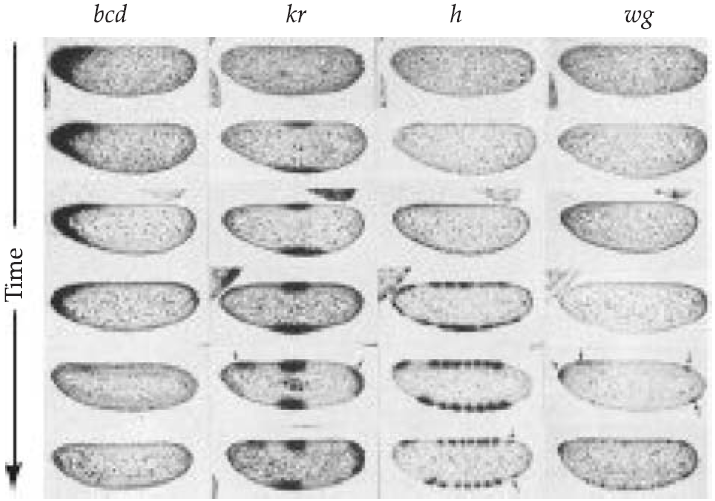
bcd: defining the anterior pole of the embryo.
kr: expressing in the middle and then at the posterior end of the embryo, defining the anterior-posterior axis.
h: delineating the domains that will eventually form the mature segmented body.
wg: further refining the organization of Individual segments.
1. Parallels between Drosophila and human Hox genes
Human Hox genes (and those of most mammals) have been duplicated twice, leading to four Independent groups (A, B, C, D), each on a distinct human chromosome.
The anterior-to-posterior pattern of Hox gene expression in both flies and mammals follows the 5’-to-3’ orientation of these genes on their respective chromosomes
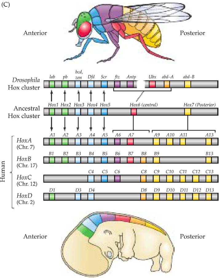
2. Hox gene expression in developing nervous system
Hox gene expression coincides with, or even precedes, the formation of morphological features–the various bends, folds, and constrictions that underlie the progressive regionalization of the developing neural tube, particularly in the hindbrain and spinal cord.
In vertebrates, Hox gene expression does not extend into the midbrain or forebrain.
Regional differences in expression of other transcription factors such as Dlx and Pax are seen in these subdivisions prior to and during the morphogenetic events that define them.
三、Molecular basis of neural induction
Neural stem cells in the early neural plate and tube, and subsequently within each nascent brain region, must acquire instructions that establish their capacity to make nerve cells specific to each region.
These instructions come from neighboring cells or tissues.
To Define
To define: Using a variety of classic experiments based on the removal or transfer of embryonic tissues
What happened to the cells that are moved into a new place:
Acquiring the identity of the new region in which they are placed (thus receiving instructions based on their new location).
Retain an identity that reflects their original position (and thus possess immutable instructions from their original location).
What happened to the old place after the cells are removed:
Compensated by local cell proliferation, causing little noticeable disruption of subsequent development (a process referred to as embryonic regulation).
Disrupting subsequent development.
What happened to the new place after the cells are moved in:
Complete change of local developmental program.
Interactions between cells in adjacent germ layers are essential for regional and cellular identity in the developing embryo.
Generally, generation of cell identity and diversity results from the spatial and temporal control of different sets of genes by endogenous signaling molecules.
Most of these molecular signals are secreted by one embryonic cell class or tissue and then diffuse through extracellular space to act on an adjacent cell class or tissue.
Neural induction: apposition of the notochord to the overlaying ectoderm is essential for establishing neuroectodermal stem cells and thus the entire nervous system.
Major inductive signaling pathways in vertebrate embryos
The embryonic structures (including the notochord, floorplate, roofplate, and neuroectoderm itself, as well as adjacent mesodermally derived tissues such as somites) that are critical for the induction and patterning of the central nervous system, all secrete these inductive signals.
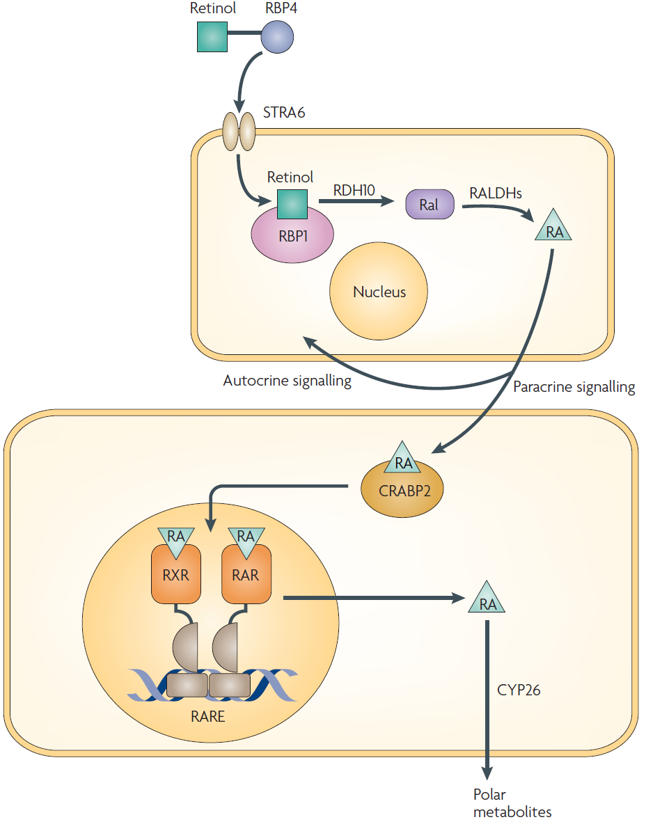
1. Retinoic acid (RA,视黄酸)
RA: One of the first inductive signals to be identified; a derivative of vitamin A.
RA signaling drives cellular differentiation, regulating the transitions between various classes of neural stem cells leading up to their terminal division for neurogenesis.
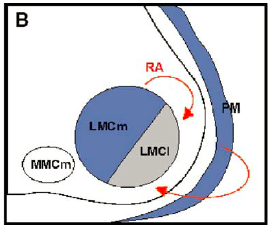
RA in motor neuron development: Sources? Mechanisms?
- Sources: From PM and LMCm
2. Retinoids (类维生素A)
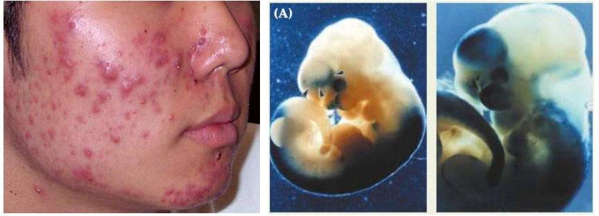
Retinoids: inductive signal and teratogen(畸胎原)
In the early 1930s, investigators noticed that vitamin A deficiency during pregnancy in animals led to a variety of fetal malformations. The most severe abnormalities affected the developing brain, which was often grossly malformed.
Experimental studies yielded the surprising finding that excess vitamin A caused similar defects.
Retinoids include the alcohol, aldehyde, and acid forms of vitamin A (retinol, retinal, and retinoic acid, respectively), are teratogenic(teratogenesis is the term for birth defects induced by exogenous agents)
Accutane (the generic name for 13-cis-retinoic acid) was used to treat severe acne. Women who used this drug during pregnancy had an increased number of spontaneous abortions and children born with a range of birth defects.
3. Fibroblast growth factor (FGF, 成纤维细胞生长因子)
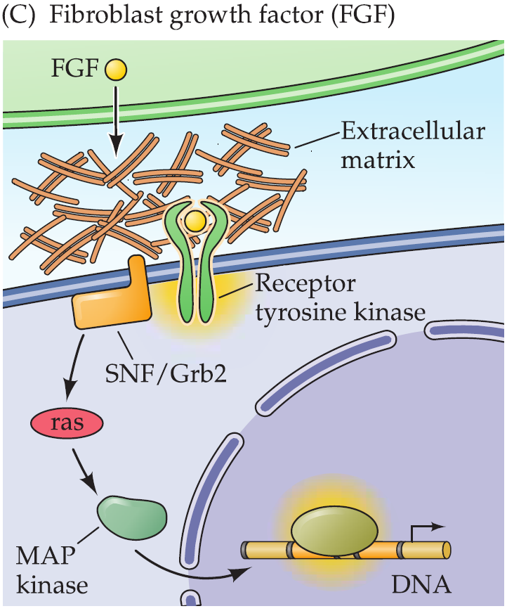
22 FGF ligands and the same receptor tyrosine kinase.
FGF8: a particularly important regulator of forebrain and midbrain development.
FGFs from the pre-somitic mesoderm (from which somites form) regulate spinal cord neurogenesis.
- By regulating the gene expression
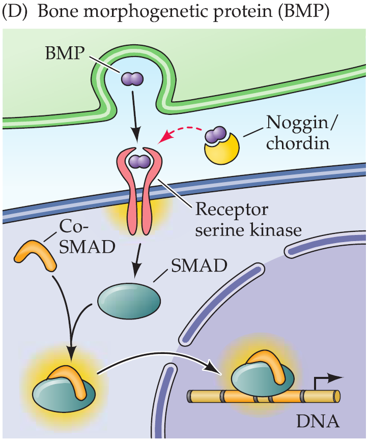
4. Bone morphogenetic proteins (BMPs, 骨塑型蛋白)
The various BMPs play roles in the initial specification of the neural plate as well as the subsequent differentiation of the dorsal part of the spinal cord and hindbrain and the cerebral cortex.
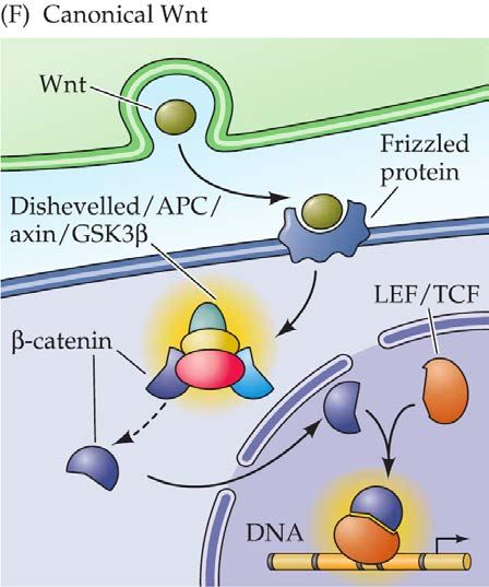
5. Wnt
The noncanonical(非经典的) pathway (planar cell polarity, PCP): featured during early nervous system morphogenesis; regulating cell movements necessary for lengthening the neural plate.
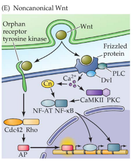
The canonical pathway: influencing cell proliferation, adhesion, and differentiation after the initial morphogenesis of the nervous system is complete
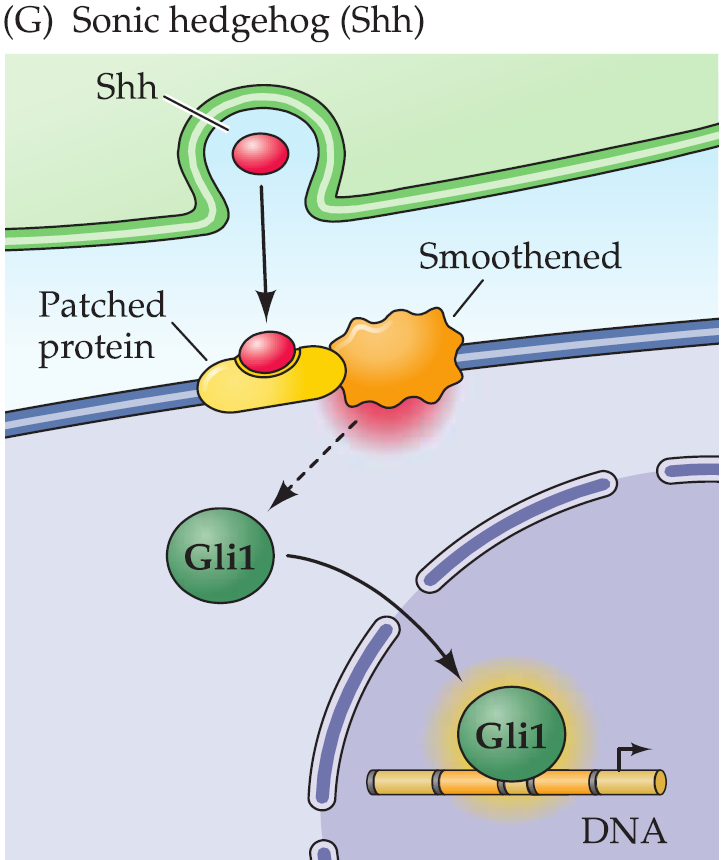
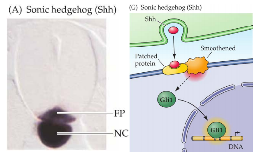
6. Sonic hedgehog (Shh, 音猬因子)
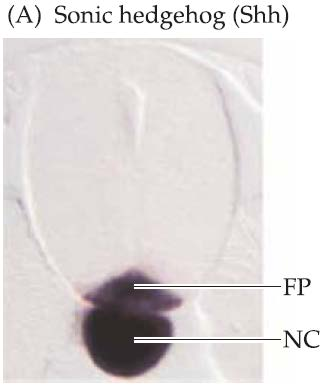
Particularly important for two phases of neural development:
Closing the neural tube, esp. the anterior midline.
Establishing the identity of neurons, esp. motor neurons in spinal cord and hindbrain.
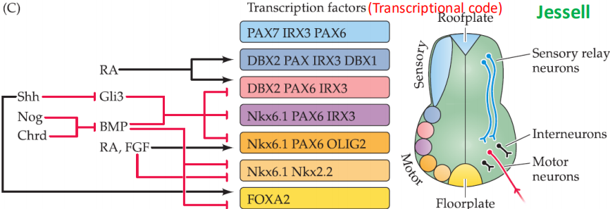
Integrated inductive signals specify neural identity
Transcriptional code: Combination of different traanscription factors
四、Neurogenesis 神经发生
神经发生是指神经元的生成。神经发生是有神经元干细胞增殖分化而成,大部分的神经发生是在胎儿发育阶段,神经发生负责将神经元密布于大脑中。
The mature human brain contains about 100 billion neurons and many more glia cells which are all generated in only a few months from a small population of precursor cells.
Neurogenesis begins after the initial patterning of the neural tube is complete, and finishes (a few exceptions) before birth.
A developing human generates about 250,000 new neurons per minute during the peak of cell proliferation.
Then precursor cells mostly disappear and, in most brain regions, few if any new neurons can ve added to replace those lost by age or injury.
The initial differentiation of neurons and glia
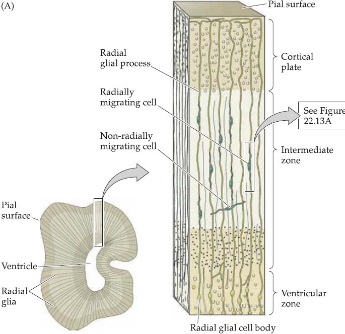
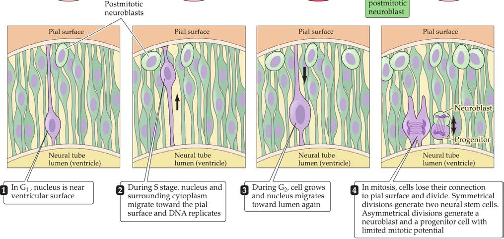
Neural precursor cells undergo mitosis within the ventricular zone (心室区).
Symmetrical division generates two new stem cells. [Self-renew]
- dividing relatively slowly and renewing themselves indefinitely.
Asymmetrical division generates a postmitotic neuroblast (有丝分裂后神经母细胞) and a progenitor with limited mitotic potential.
dividing more rapidly but with a limited capacity for division over time.
transit amplifying cells: a transitional form between stem cells and differentiated neurons, and account for the amplification in numbers of differentiated cells
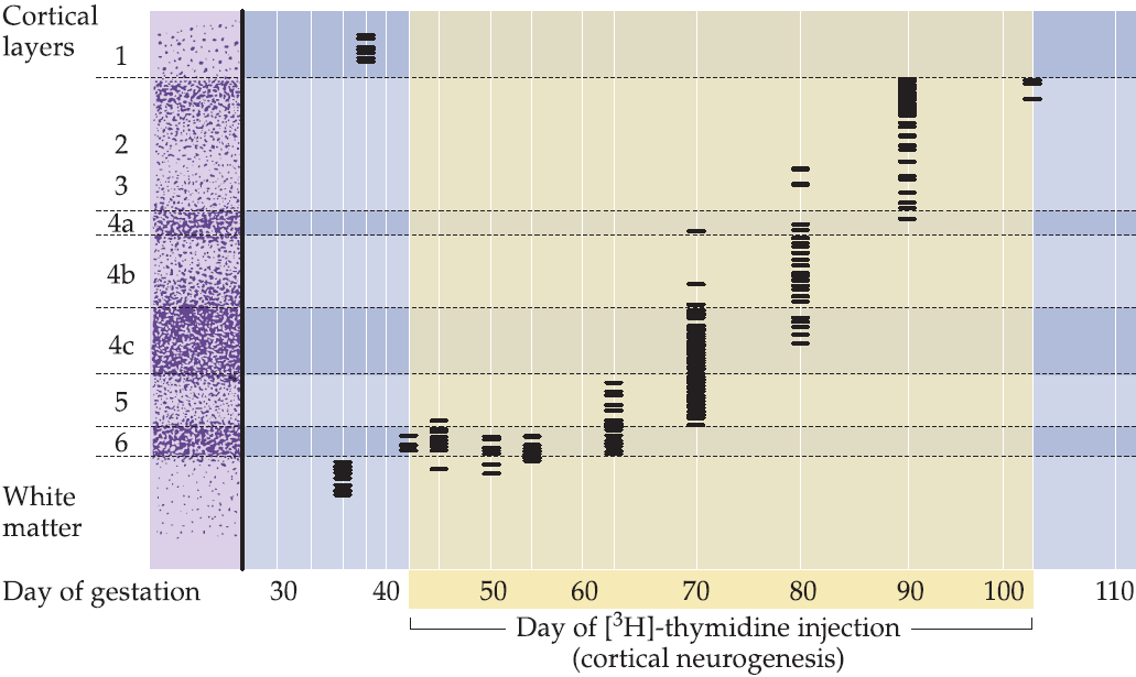
Neurogenesis in cerebral cortex(大脑皮层)
Most neurons of the six cortical layers are generated in an inside-out manner:
- Each layer consists of a cohort of cells “born” at a distinct time.
- The first born cells are eventually located in the deepest layers.
- Except layer 1
- Later generations of neurons migrate radially from the site where they are born, traveling through the older cells and coming to lie superficial to them.
- Early born neurons in the lower cortical layers express transcription factors distinct from those of the later-born neurons in the upper cortical layers.
- In most regions of the central nervous system where neurons are arranged into layered or laminar structures (the hippocampus, cerebellum, superior colliculus, and retina), there is a systematic relationship between the layers, time of cell origin, and additional molecular properties, including transcription factor expression
Molecular regulation of neurogenesis
Neural stem cell decisions to generate either additional stem cells or postmitotic neurons**: Delta-Notch** signaling
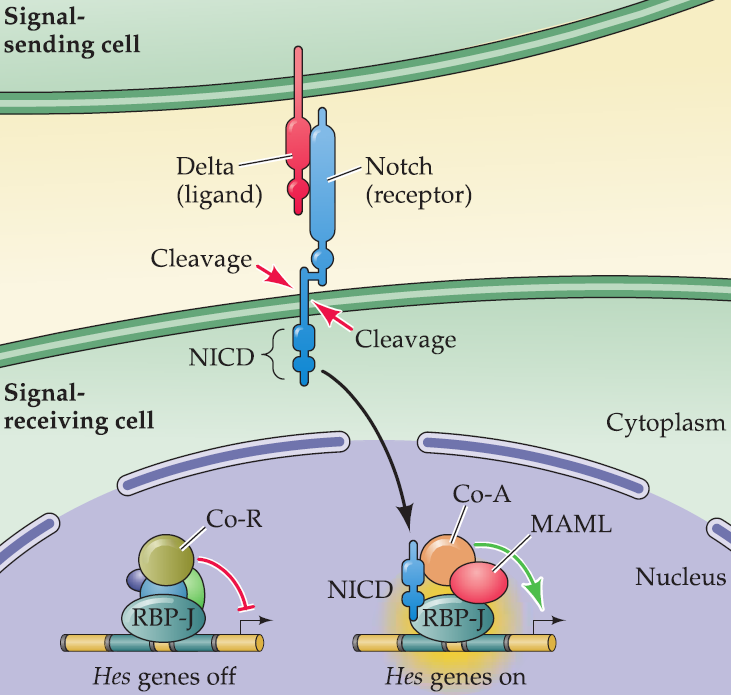
- NICD is cleaved and travels to the nucleus, then the cell can generate bHLH neurogenic factors
Molecular and genetic disruptions of early neural development
Molecules involved in neural induction, neurogenesis, and generation of neuronal diversity: inductive signaling molecules and related signaling proteins
- Maternal dietary insufficiency of substances such as folic acid can disrupt embryonic neural tube formation.
- Cause the neuro tube can not close
- Excessive maternal intake of vitamin A–the metabolic precursor of retinoic acid–can impede neural tube closure and differentiation, or later aspects of neuronal differentiation.
- Embryonic exposure to a variety of other drugs such as alcohol and thalidomide can also elicit pathological differentiation of the embryonic nervous system by providing or blocking inductive signals at in appropriate times or places.
- Because the consequences of disordered neural induction are so severe, pregnant women are advised to avoid virtually all drugs and dietary supplements except those specifically prescribed by a physician, especially during the first trimester
Triple Jeopardy (危险): diseases associated with Sonic Hedgehog
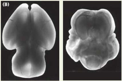
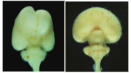
Holoprosencephaly(前脑无裂畸形): disrupting the initial morphogenesis of two distinct cerebral hemispheres.
Medulloblastoma: cancerous transformation of cerebellar granule neuron precursor cells.
Basal cell carcinoma: the most prevalent cancer of the skin (basal epidermal cells (基底表皮细胞))
1. Holoprosencephaly
1 in every 16,000 live births and the most common malformation of the mammalian forebrain known.
Compromising the normal separation of the two hemispheres of the forebrain and disrupting development of midline facial structures.
The most severe form: failure of the eye primordium to split into two bilaterally symmetrical fields and the subsequent development of a single eye (cyclopia).
A small but consistent cases are associated with deletions or missense mutations in SHH on chromosome 7. Supported by studies in mice and zebrafish
Neuronal migration in PNS
In the developing nervous system, migration brings different classes of neurons together so that they can interact, and insures the final position of many postmitotic neurons.
The developing neuron must be in the right place at the right time so that it can be properly integrated into a functional circuit that can mediate behavior.
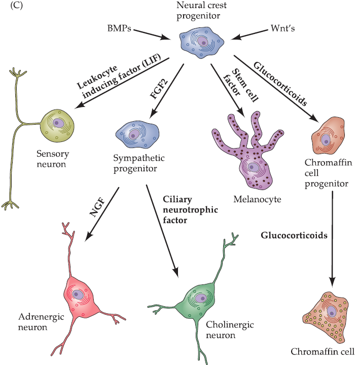
Migration and differentiation of neural crest cells from the neural tube to the periphery of the embryo.
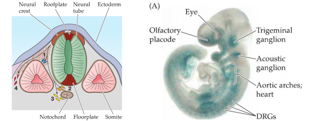
1. Neural crest migration 神经嵴迁移
They begin as neuroepithelial cells, with all of the intercellular junctions and adhesive interactions that keep epithelial cells in place.
To move, they need undergo an epithelial-to-mesenchymal transition. (mesenchymal cells being loosely packed and tending to migrate freely)
How? They express bHLH family members Snail1 and Snali2, which repress expression of intercellular junctional proteins and epithelial adhesion molecule.
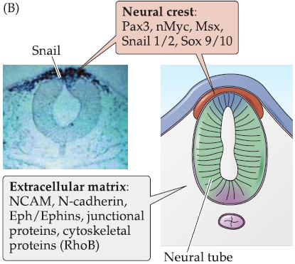
Neural crest cells are largely guided along distinct migration pathways by signals from non-neural peripheral structures such as somites and other rudimentary musculoskeletal or visceral tissues.
The signals along these pathways can be secreted molecules such as neurotropic factors and peptide hormone growth factors, cell surface ligands and receptors such as N-cadherin, NCAM or ephreceptors and ephrin ligands, or extracellular matrix molecules such as laminin and fibronectin.
Neuronal migration in CNS
Minority of neurons and glia: using existing axon pathways as migratory guides.
Most prominent form: guided by glial cells—radial glia.