L02 Microscopy
一、Microscopy
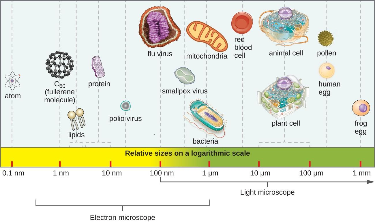
Why microorganisms are good experimental tools?
- Easy culture and manipulate, store…
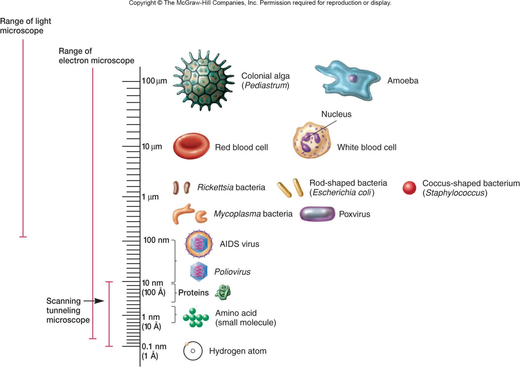
The Light Microscope
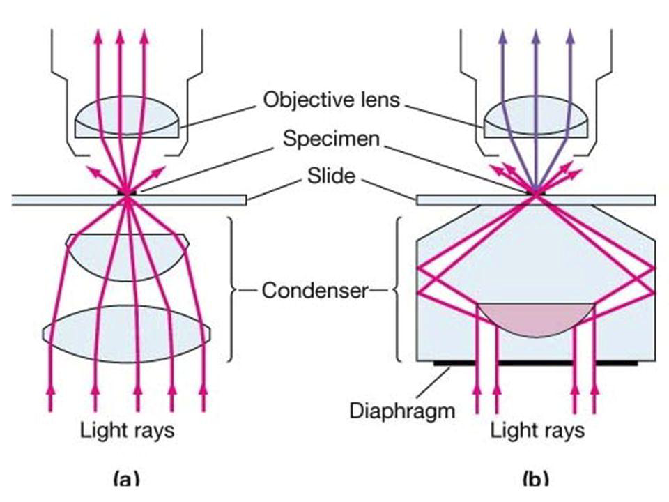
1. The Bright-Field Microscope 明场显微镜
- Produces a dark image against a brighter background
- Has several objective lenses
- Total magnification
- the ocular lenses and the objective lenses
The specimen is dark and contrasted by the surrounding bright viewing field.
By using an aperture diaphragm for contrast, past a certain point, greater contrast adds distortion(失真). However, employing an iris diaphragm will help compensate for this problem.
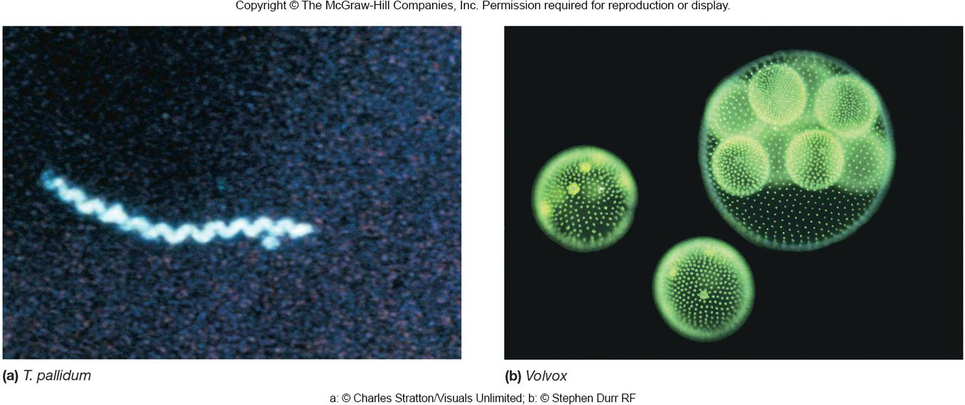
2. The Dark-Field Microscope 暗场显微镜
Image is formed by light reflected or refracted by specimen
Produces a bright image of the object against a dark background
Used to observe living, unstained preparations
internal structures
identify bacteria
Produces a bright image of the object against a dark background
Used to observe living, unstained preparations
- internal structures
- identify bacteria
3. The Phase-Contrast Microscope 相差显微镜
Converts differences in refractive index/cell density into detected variations in light intensity
Some light rays from hollow cone of light passing through unstained cell slowed/out of phase (dark against bright background)
Excellent way to observe living cells
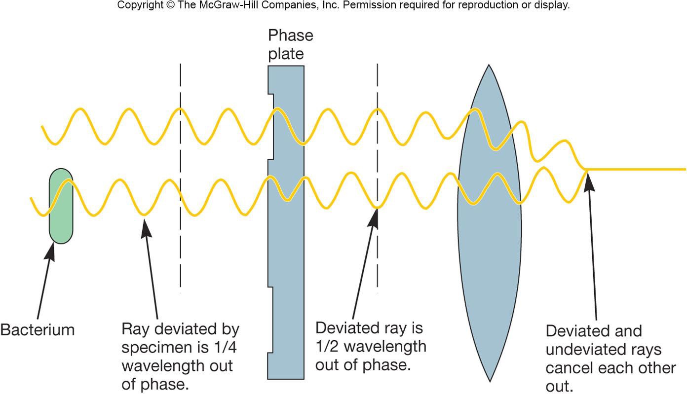
As this cone of light passes through a cell, some light rays are bent due to variations in density and refractive index within the specimen, and are retarded by about 1/4 wavelength. The deviated light is focused to form an image of the object.
Excellent way to observe living cells.
4. The Differential Interference Contrast Microscope (DIC) 差分干涉对比显微镜
Creates image by detecting differences in refractive indices and thickness of different parts of specimen
Excellent way to observe living cells
- live, unstained cells appear brightly colored and three-dimensional
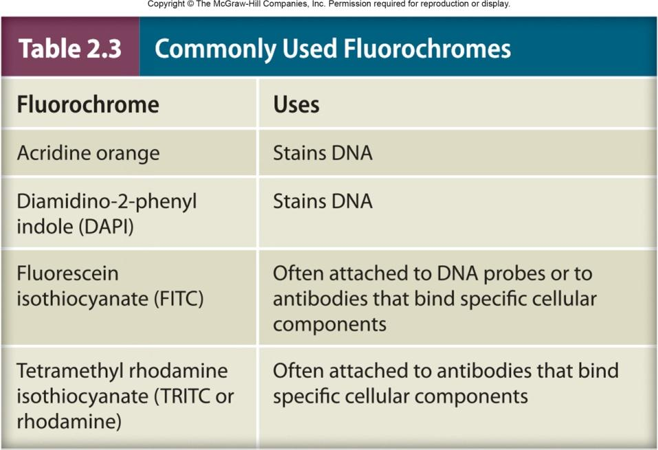
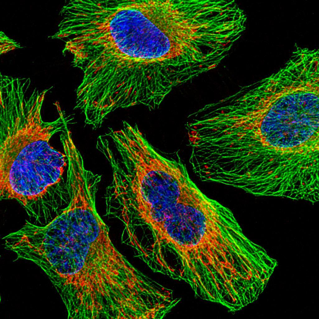
5. The Fluorescence Microscope 荧光显微镜
Specimens usually stained with fluorochromes(荧光染料)
Shows a bright image of the object resulting from the fluorescent light emitted by the specimen
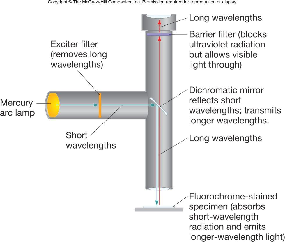
Essential tool in microbiology
- fluorochrome-labeled probes, such as antibodies, for identification of unknown pathogens
- localization of specific proteins in cells
6. Confocal Microscopy 共聚焦显微镜
共聚焦显微成像技术(英语:Confocal microscopy)是一种利用逐点照明和空间针孔调制来去除样品非焦点平面的散射光的光学成像手段,相比于传统成像方法可以提高光学分辨率和视觉对比度。
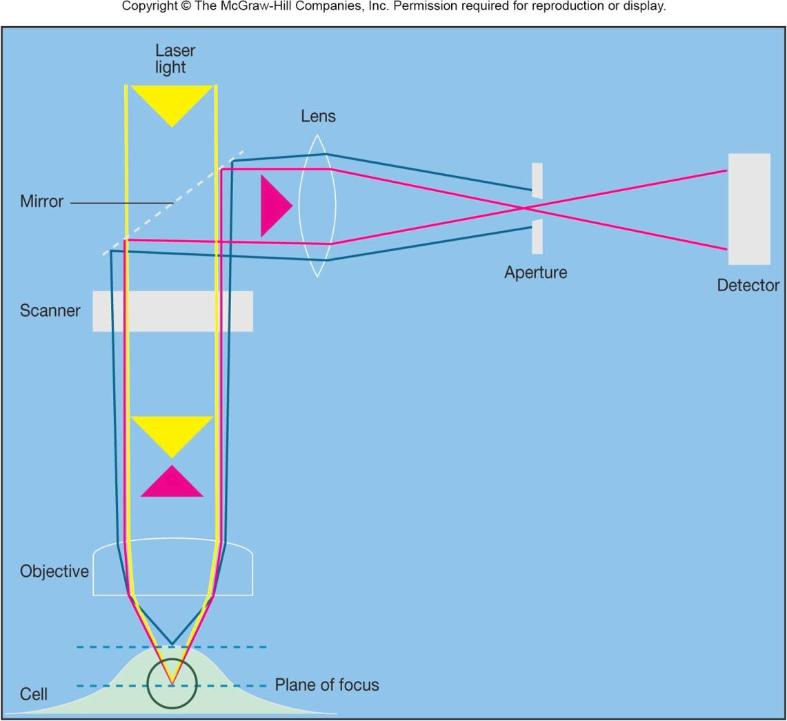
Confocal scanning laser microscopy (CLSM) creates sharp, composite 3D image of specimens by using laser beam, aperture to eliminate stray light, and computer interface.
二、Preparation and Staining of Specimens
- Increases visibility of specimen
- Accentuates(强调) specific morphological features
- Preserves specimens
Fixation
Preserves internal and external structures and fixes them in position
Organisms usually killed and firmly attached to microscope slide
1. Heat fixation
routinely used with bacteria and archaea
- preserves overall morphology(形态学) but not internal structures
In order to making the cells attach firmly to the slide
2. Chemical fixation
used with larger, more delicate organisms
- protects fine cellular substructure and morphology
Dyes and Simple Staining
Dyes
make internal and external structures of cell more visible by increasing contrast with background
| Type of Dye | Examples | Characteristics |
|---|---|---|
| Basic Dye | Methylene blue, basic fuchsin, crystal violet, safranin, malachite green | Have positively charged groups; bind to negatively charged molecules such as nucleic acids, many proteins, and the surfaces of bacterial and archaeal cells |
| Acidic Dye | Eosin, rose bengal, and acid fuchsin—possess groups such as carboxyls (—COOH) and phenolic hydroxyls (—OH). | In their ionized form, have a negative charge and bind to positively charged cell structures |
Classification of Staining
(1) Simple Staining
Simple stains
a single stain is used
use can determine size, shape, and arrangement of bacteria
(2) Differential Staining
Differential stain used to detect presence or absence of structures
- endospores, flagella, capsules
Divides microorganisms into groups based on their staining properties
- e.g., Gram stain
- e.g., acid-fast stain
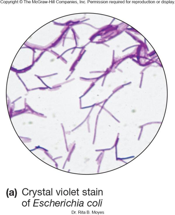
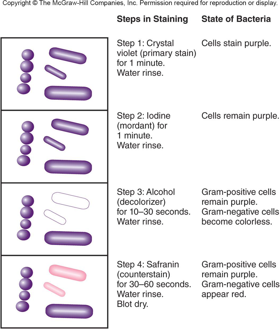
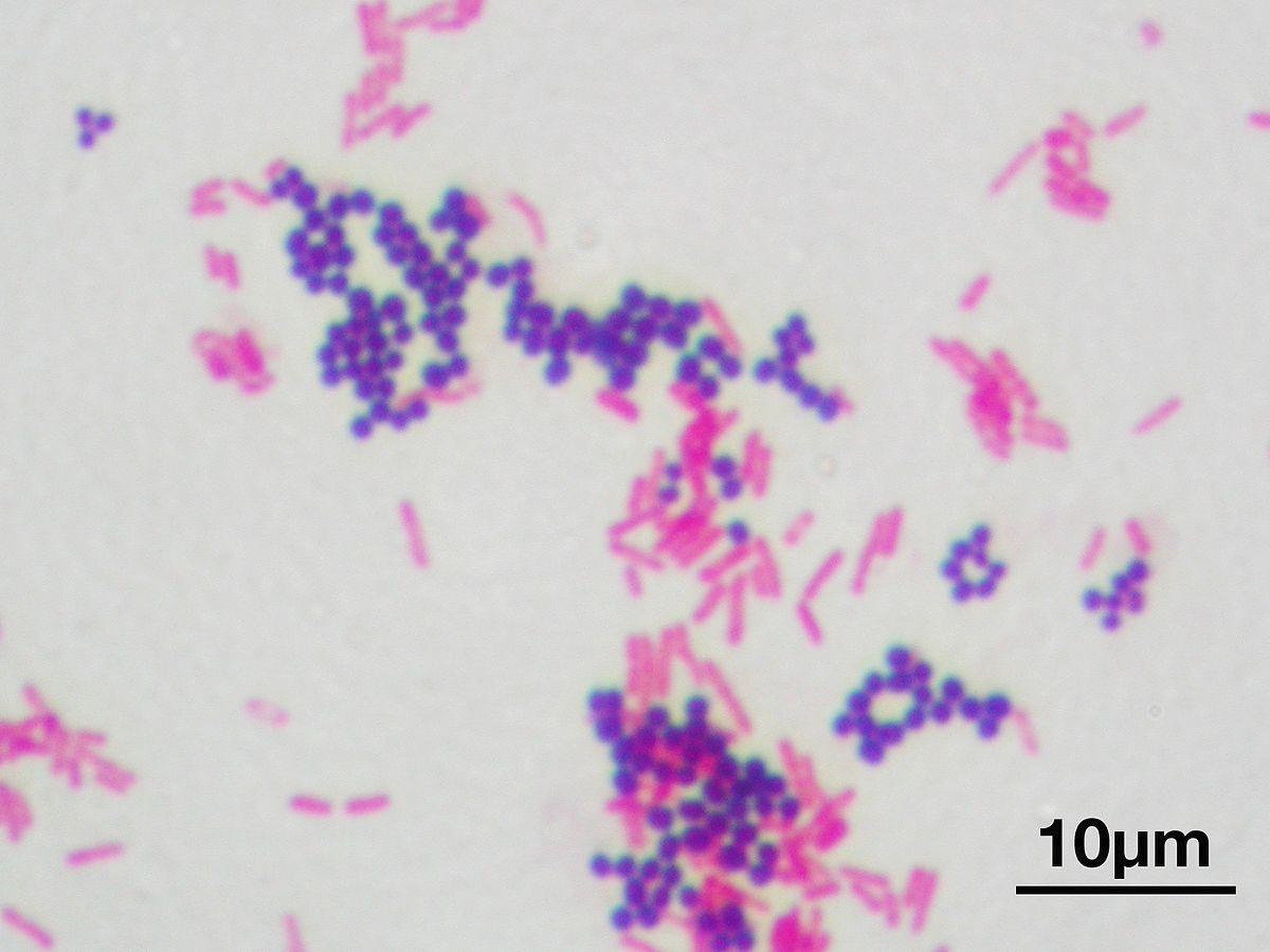
1. Gram Staining 革兰氏染色
Most widely used differential staining procedure
Divides bacteria into two groups, Gram-positive and Gram-negative, based on differences in cell wall structure
革兰氏染色(Gram Staining)是用来鉴别细菌的一种方法,细菌细胞壁上的主要成份不同,利用这种染色法,可将细菌分成两大类,即革兰氏阳性菌与革兰氏阴性菌(Gram-positive and Gram-negative)。
这种染色法是由一位丹麦医生汉斯·克里斯蒂安·革兰(Hans Christian Gram,1853年-1938年)于1884年所发明,最初是用来鉴别肺炎球菌与克雷白氏肺炎菌之间的关系。
Crystal violet (primary stain) $\rarr$ Iodine(mordant) $\rarr$ Alcohol (decolorizer) $\rarr$ Safranin (counterstain)
decolorizer时间长的话,counterstain 后 gram-positive cells 也会变成粉色
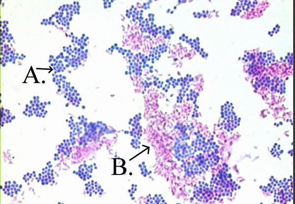
2. Acid-Fast Staining 抗酸细菌染色法
Particularly useful for staining members of the genus Mycobacterium
- e.g., Mycobacterium tuberculosis - causes tuberculosis
- e.g., Mycobacterium leprae 一 causes leprosy
high lipid content in cell walls (mycolic acid) is responsible for their staining characteristics
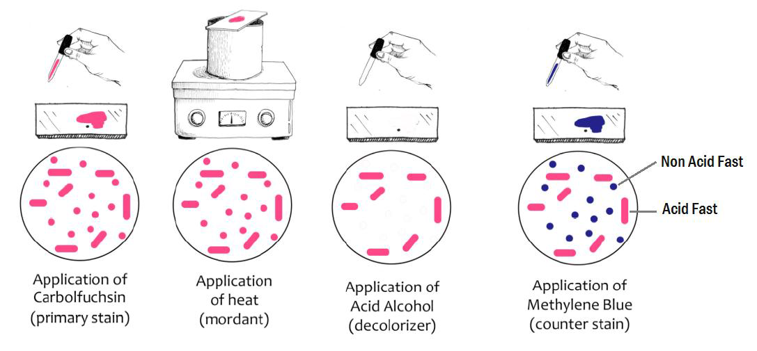
ACID-FAST (adjective) The adjective ACID-FAST has 1 sense that not easily decolorized(脱色) by acid solutions; pertains to micro-organisms (especially the tubercle bacillus that causes tuberculosis) Familiarity information: ACID-FAST used as an adjective is very rare.
high lipid content in cell walls (mycolic acid) is responsible for their staining characteristics
3. Staining Specific Structures
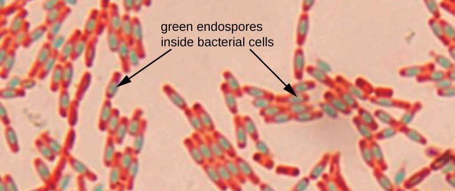
Endospore staining
bacterial endospore is one color and vegetative cell(营养细胞) is a different color
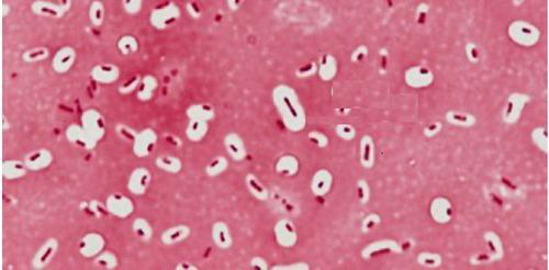
Capsule stain 荚膜染色
Capsule stain used to visualize polysaccharide capsules surrounding bacteria
- negative stain - capsules may be colorless against a stained background
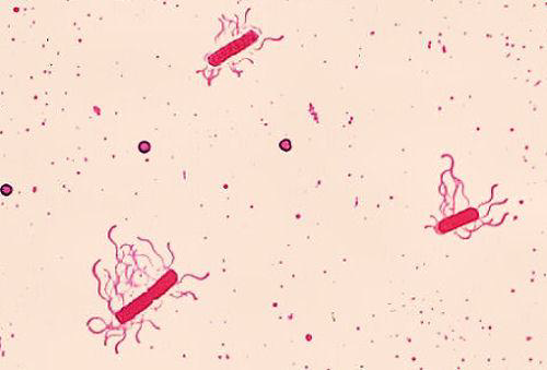
Flagella staining 鞭毛染色
mordant applied to increase thickness of flagella
三、Electron Microscope
Electrons replace light as the ‘illuminating’ beam
Wavelength of electron beam is much shorter than light, resulting in much higher resolution
Allows for study of microbial morphology in great detail
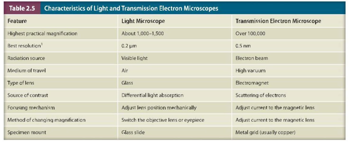
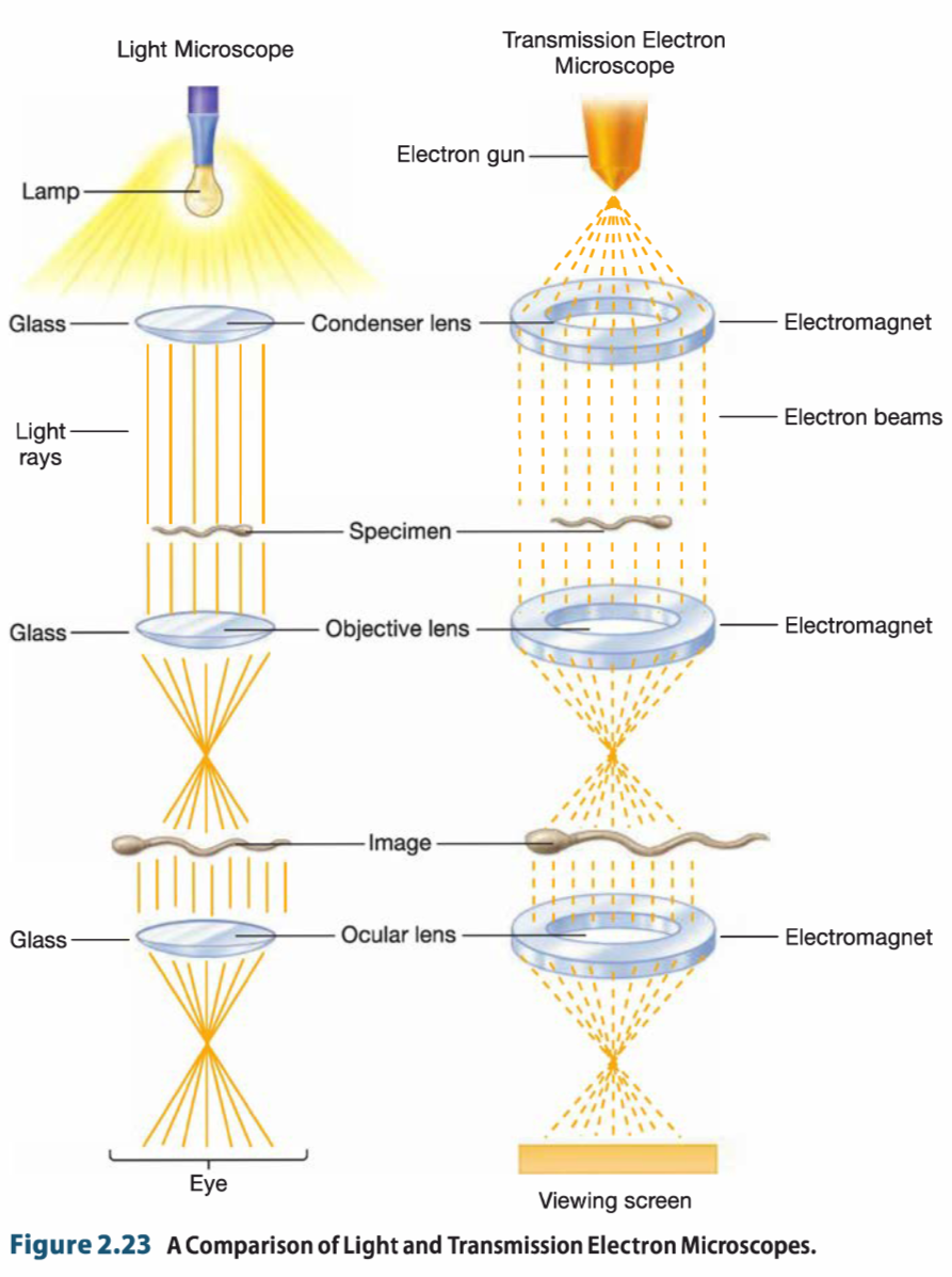

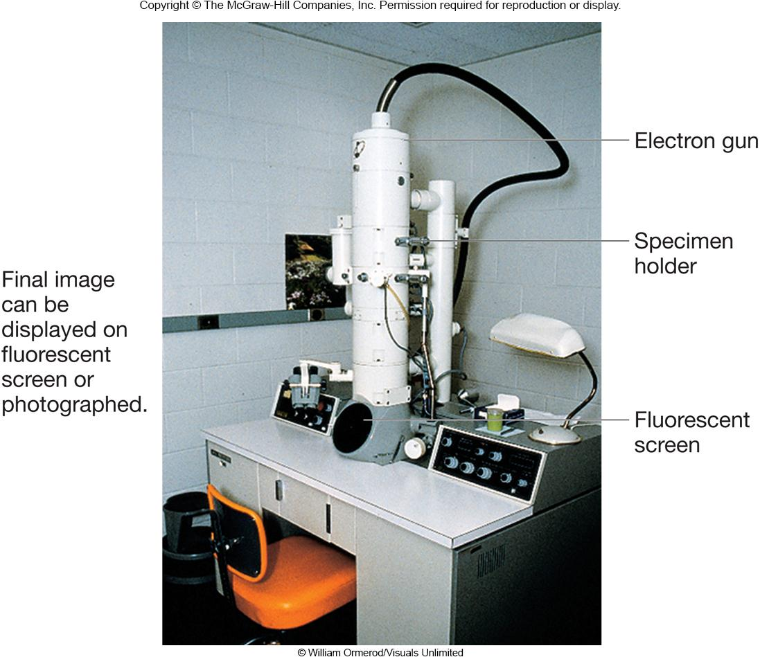
Transmission Electron Microscope 透射电子显微镜(TEM)
Transmitted electrons are under vacuum which reduces scatter and are used to produce clear image
Electrons scatter when they pass through thin sections of a specimen
Denser regions in specimen scatter more electrons and appear darker
A heated tungsten(钨) filament in the electron gun generates a beam of electrons that is focused on
the specimen by the condenser.
The column containing the lenses and specimen must be under high vacuum to obtain a clear image because electrons are deflected by collisions with air molecules.
当电子碰到样本的时候,就会发生碰撞,最终导致无法到达下方的Viewing Screen,从而形成一片”electron dense“
1. Specimen Preparation for EM
Analogous to procedures used for light microscopy
For transmission electron microscopy, specimens must be cut very thin
Specimens are chemically fixed and stained with electron dense materials, such as heavy metals, that differentially scatter electrons
- 样本厚度:Specimens must be viewed in a vacuum because elec trons are deflected by air molecules. Only extremely thin slices (20 to 100 nm) of a specimen can be viewed because electron beams are easily absorbed and scattered by solid matter.
因此对于样本的第一个要求是非常薄 - 染色:电子的scatter(散射)取决于the density (atomic number) of atoms in the specimen,而普通的样本组成元素为C、H、O、N,electron scattering is fairly constant throughout an unstained cell or virus.
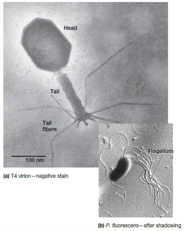
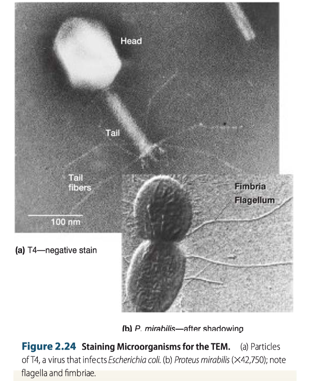
Therefore specimens are prepared for observation by soaking thin sections with solutions of heavy metal salts such as lead citrate(柠檬酸铅) and uranyl acetate(醋酸铀酰). - negative staining(负染色):In this technique, the background is stained, leaving the actual specimen untouched, and thus visible. This contrasts with ‘positive staining’, in which the actual specimen is stained. 即对背景染色,使得样本visible. Negative staining is an excellent way to study the structure of virus particles, bacterial gas vacuoles, and other similar objects.
- Shadowing:In shadowing, a specimen is coated with a thin film of platinum or other heavy metal by evaporation at an angle of about 45° from horizontal so that the metal strikes the microorganism on only one side. In one commonly used imaging method, the area coated with metal appears dark in photographs, whereas the uncoated side and the shadow region created by the object are light. This technique is particularly useful in studying virus particle morphology, bacterial and archaeal flagella, and DNA.
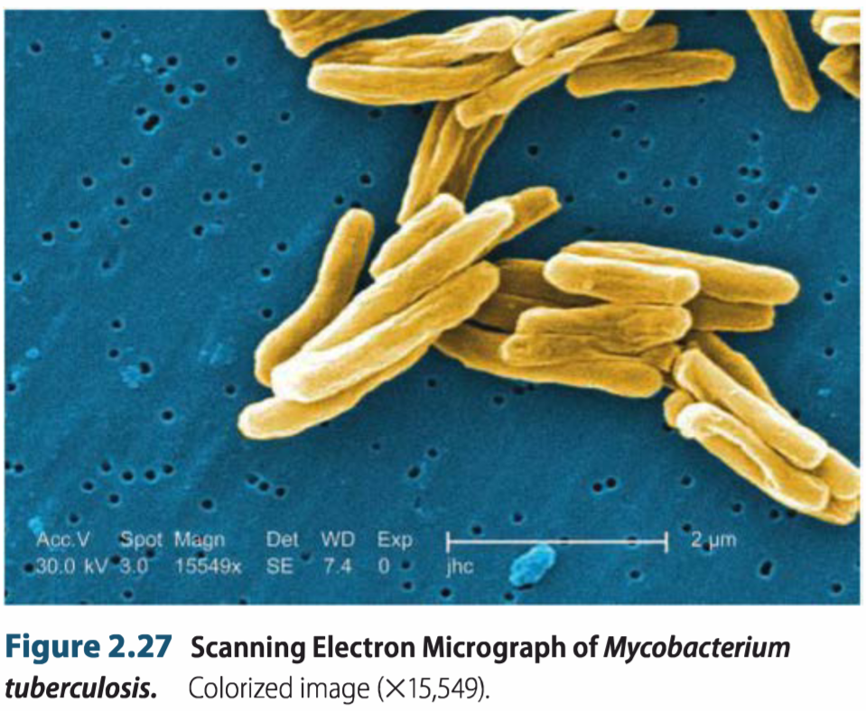
The Scanning Electron Microscope 扫描电子显微镜(SEM)
Uses electrons excited from the surface of a specimen to create detailed image
Produces a realistic 3D image of specimen’s surface features
Can determine actual in situ location of microorganisms in ecological niches
Transmission electron microscopes form an image from radiation that has passed through a specimen.
The scanning electron microscope (SEM) produces an image from electrons released from atoms on an object’s surface.
SEM used to observe the SURFACE structure
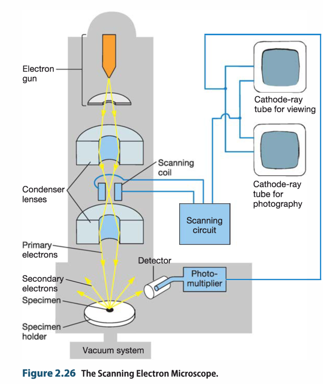
工作原理:When the beam strikes a particular area, surface atoms discharge a tiny shower of electrons called secondary electrons, and these are trapped by a detector. Secondary electrons entering the detector strike a scintillator, causing it to emit light flashes that a photo multiplier converts to an electrical current and amplifies. The sig nal is sent to a cathode-ray tube and produces an image that can be viewed or photographed.
Electron Cryotomography 电子冷冻断层扫描(冷冻电镜)
Rapid freezing technique provides way to preserve native state of structures examined in vacuum
Images are recorded from many different directions to create 3-D structures
Provides extremely high resolution images of
- cytoskeletal elements, magnetosomes, inclusion bodies, flagellar motors, viral structures
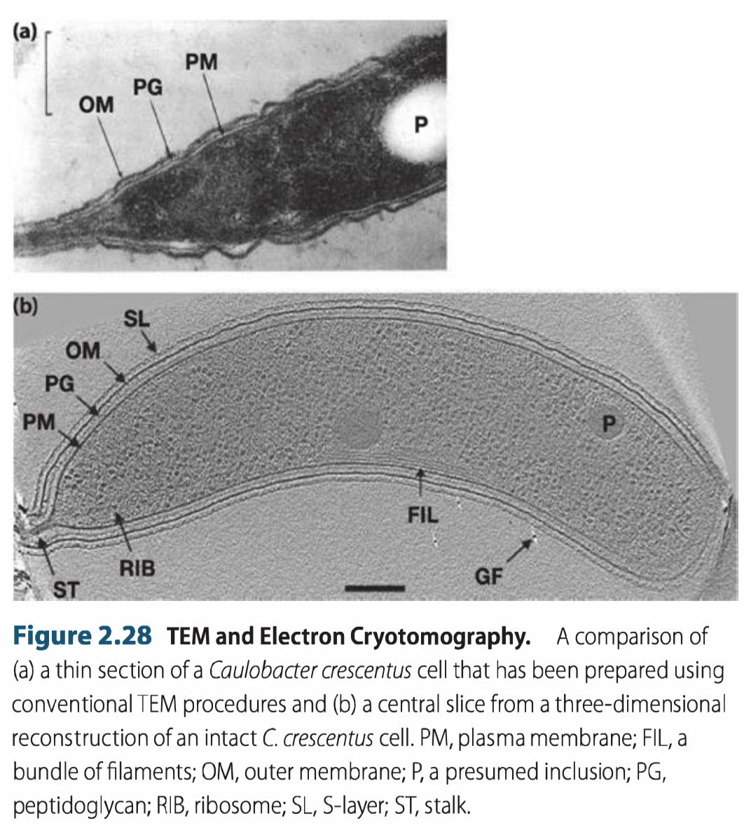
Cryo- refers to sample preparation and visualization.
Samples are prepared by rapidly plunging the specimen into an extremely cold liquid (e.g., ethane); the sample is kept frozen while being examined.
Rapid freezing of the sample results in the formation of vitreous(玻璃状的) ice rather than ice crystals. 这种玻璃状的冰可以保持native state of structures and immobilizes(固定) the specimen.
The object is viewed from many directions, referred to as a tilt series
Scanning Probe Microscopy 扫描探针显微镜
扫描探针显微镜(Scanning probe microscopy,SPM)是所有机械式地用探针在样本上扫描移动以探测样本影像的显微镜的统称。其影像分辨率主要取决于探针的大小〔通常在奈米的范围〕。
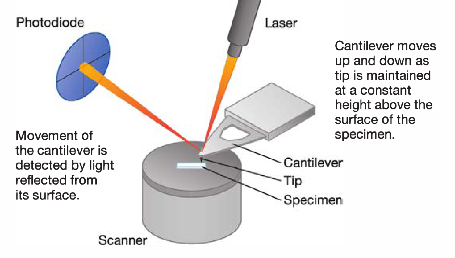
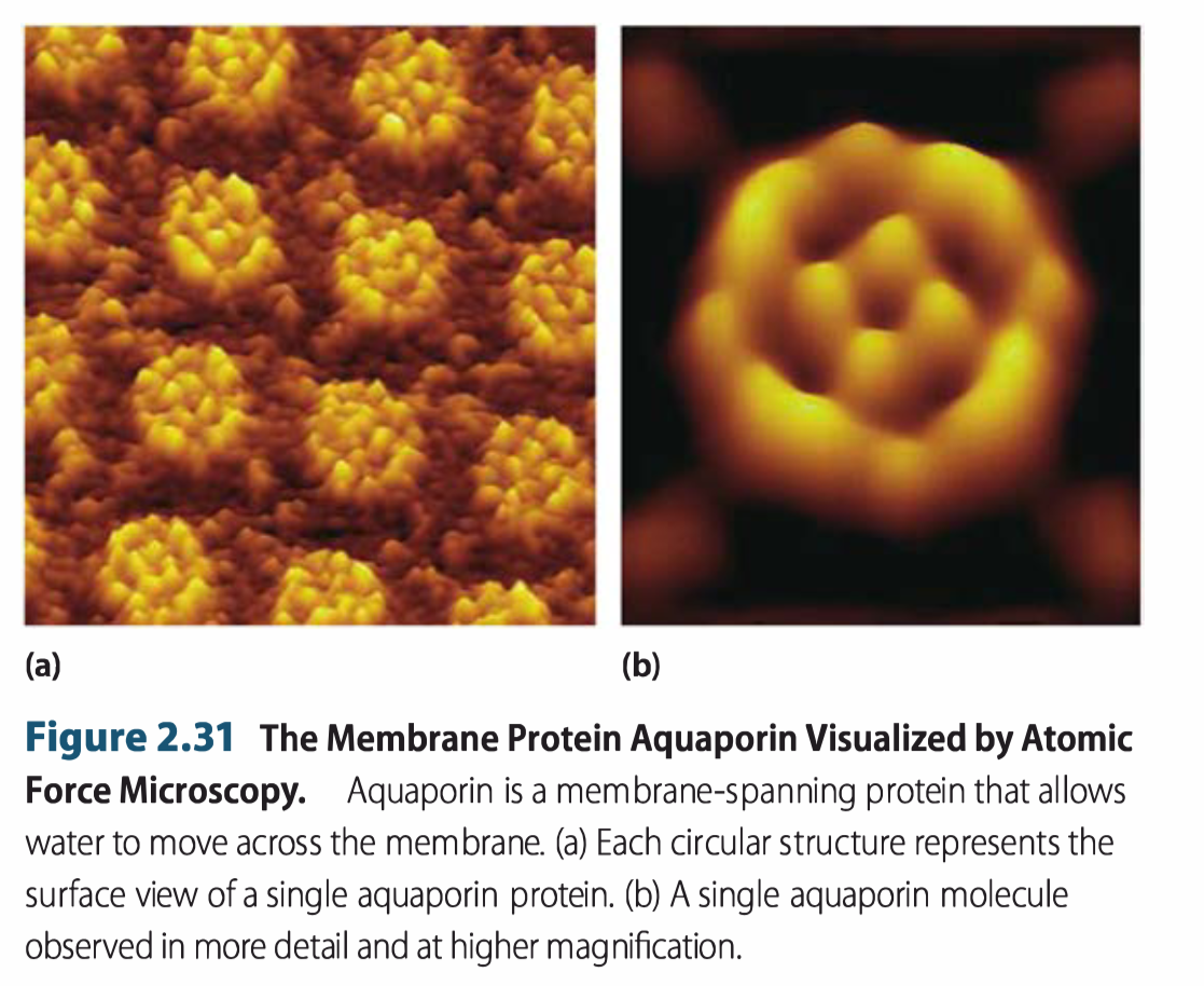
1. Atomic Force Microscopy(原子力显微镜)
Atomic force microscope
- sharp probe moves over surface of specimen at constant distance
- up and down movement of probe as it maintains constant distance is detected and used to create image
- magnification 100 million times, can view atoms on surface of a solid
原子力显微镜(atomic force microscope,简称AFM),也称扫描力显微镜(scanning force microscope,SFM)是一种纳米级高分辨的扫描探针显微镜,优于光学衍射极限1000倍。
AFM是在纳米尺度操作材料,及其成像和测量最重要的工具。信息是通过微悬臂感受和悬臂上尖细探针的表面的“感觉”来收集的,而压电组件可以控制样品或扫描器非常精确的微小移动,用导电悬臂(cantilever)和导电原子力显微镜附件则可以测量样品的电流偏压;更高级的仪器则可以测试探针上的电流来测试样品的电导率或下表面的电子的移动,不过这种测试是非常艰难的,只有个别实验室报道了一致的数据。利用微悬臂感受和放大悬臂上尖细探针与受测样品原子之间的作用力,从而达到检测的目的,具有原子级的分辨率。
由于原子力显微镜既可以观察导体,也可以观察非导体,从而弥补了扫描隧道显微镜的不足。