L3 Methods to Study Biophysiology
一、Methods to Study Biophysiology
Firstly we can Study the patients with brain damage
Experimental Lesion
1. Permanent Lesion
Aspiration(吸引术)
Radio frequency leision
- Using heat
Excitotoxic leision (kainic acid)
- e,g,: NMDA
Specific chemical leision
- e.g.: Colchicine (秋水仙碱), specific to dentate gyrus (齿状回)
Sham leision: ‘placebo’ leision: 对照组,在不同的实验中措施不同(只插入电极不施加或者在chemical leision的过程中给予安慰剂)
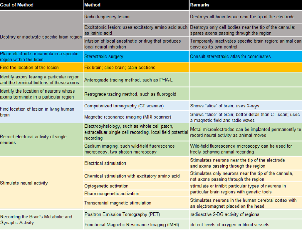
| Technique | Information Provided | Advantages | Disadvantages |
|---|---|---|---|
| Permanent Lesions | Change of behavior implies function of lesioned area | 1. Allows brain regions to be correlated with behavior 2. Easy |
May misinterpret behavior information: 1. All areas of brain interconnected 2. Redundancy of brain function |
| Aspiration | General area involved in functions | Surface lesion | Unspecific damage |
| Radio frequency lesion | General area involved in functions | Internal lesions | 1. Unspecific damage 2. Damages all cells and paths in the area |
| Excitotoxic lesion (e.g. kainic acid) | Specific area involved in functions | Selective damage destroys only cell bodies, not pathways | 1. Unspecific damage 2. Stimulates many types of neurons 3. Kills neurons that it effects when high does used |
| Specific chemical lesion (e.g. colchicine ) | Specific area involved in functions | Selective damage | Difficult to determined the does |
Radio Frequency Lesion

- Damages all cells and passing fibers in the area
- The arrows point to very small lesions produced by passing radio frequency current through the tips of stainless steel electrodes placed in the medial preoptic nucleus of a rat brain. The oblong hole in the middle of the photograph is the third ventricle. (Frontal section, cell-body stain.)
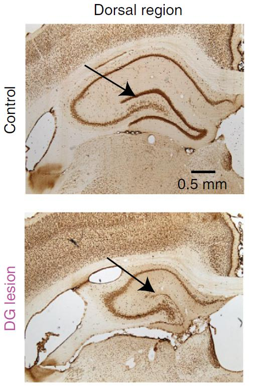
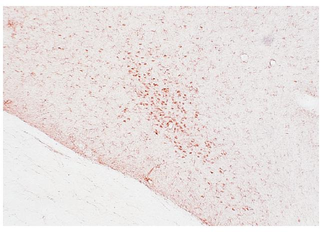
Excitotoxic Lesion

- Destroys cell bodies, but spares axons(保留轴突)
- Allows investigator to separate role of neurons from nearby axons
- Slices through the hippocampus of a rat brain.
(a) Normal hippocampus.
(b) Hippocampus with a lesion produced by infusion of an excitatory amino acid. Arrowheads mark the ends of the region in which neurons have been destroyed
Specific Chemical Lesion : Colchicine (Specific to dentate gyrus)
Region’s function can be inferred from the behaviors an animal can no longer perform after brain damage
Goal is to determine what functions are performed by different brain regions and how those functions combine to accomplish specific behaviors
- Brain function and behavior are distinct
- Brain circuits perform functions, not behaviors
- All brain regions are interconnected
2. Temporary Lesion
| Technique | Information Provided | Advantages | Disadvantages |
|---|---|---|---|
| Temporary Lesions | Change of behavior implies function of lesioned area | 1. Inhibition of functioning and behavior is reversible 2. Can be cell type specific |
1. Timing is difficult to control 2. Depends on specific device and drug, tools |
| Cooling | Specific cortex involved in functions | Reversible inactivation of the cortex by surface cooling is a powerful method for studying the function of a particular area | The same as advantages |
| Inhibition (e.g. Muscimol) | General area involved in functions | Activates GABA receptors – inhibits function of neuron in area | The same as advantages |
| Optogenetics | Specific area or cell type involved in functions | Decrease the firing rate of neurons expressing specific channels | The same as advantages |
| Pharmacogenetics 遗传药理学 | Specific area or cell type involved in functions | Decrease the firing rate of neurons expressing specific channels | The same as advantages |
- Cooling
- Inhibition
- Chemically inhibit certain area or cells
- Optogenetics
- Pharmacogenetics
3. Sham Lesion
A “placebo” procedure (“安慰剂”程序) that duplicates all the steps of producing a brain lesion except for the one that actually causes the brain damage.
- Radio frequency lesion: Insert a metal electrode without giving current
- Excitotoxic lesion: vehicle solution rather than the drug
4. Techniques
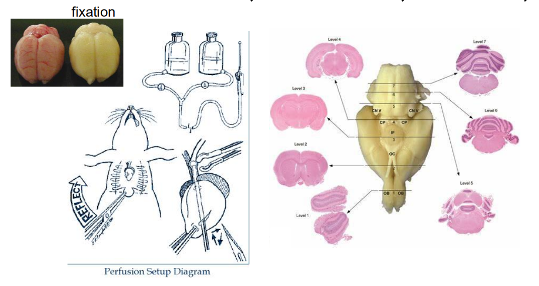
Stereotaxic surgery 立体定位手术
Brain surgery using a stereotaxic apparatus to position an electrode or cannula in a specified position of the brain.
Stereotaxis literally means “solid arrangement”; more specifically, it refers to the ability to locate objects in space
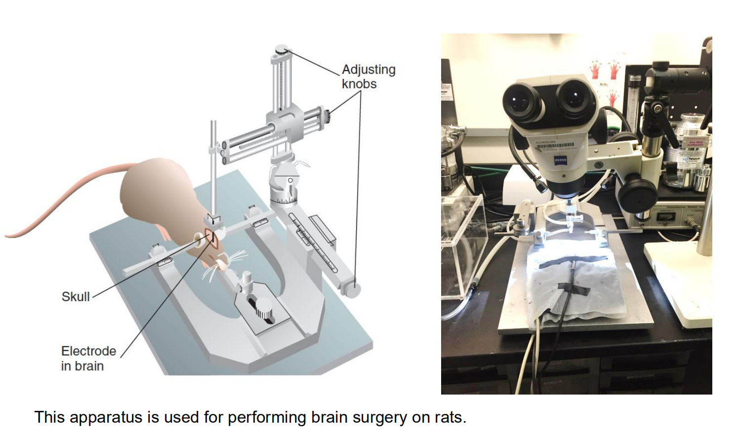
- Stereotaxic apparatus 立体定位仪
- A device that permits a surgeon to position an electrode or cannula into a specific part of the brain
- Stereotaxic atlas 立体定位图谱
- A collection of drawings of sections of the brain of a particular animal with measurements that provide coordinates for stereotaxic surgery
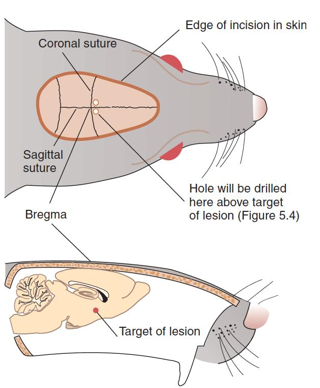
Bregma 前囟
- The junction of the sagittal and coronal sutures of the skull; often used as a reference point for stereotaxic brain surgery.
Example of stereotaxic atlas:

- This sample page from a stereotaxic atlas shows a drawing of a frontal section through the rat brain. The target (the fornix) is indicated in red.
Histological Methods
- Cardiac Perfusion 心脏灌注
- Fixation with Formalin or Paraformaldehyde (PFA)
- Section with a Microtome
- An instrument that produces very thin slices of body tissues.
- Staining
- In the late nineteenth century Franz Nissl, a German neurologist, discovered that a dye known as methylene blue would stain the cell bodies of brain tissue.
- The material that takes up the dye, known as the Nissl substance, consists of RNA, DNA, and associated proteins located in the nucleus and scattered, in the form of granules, in the cytoplasm.
- Microscopy
- Light microscopy
- Electron microscopy: A microscope that provides three-dimensional information about the shape of the surface of a small object.

Tracing Neural Connections
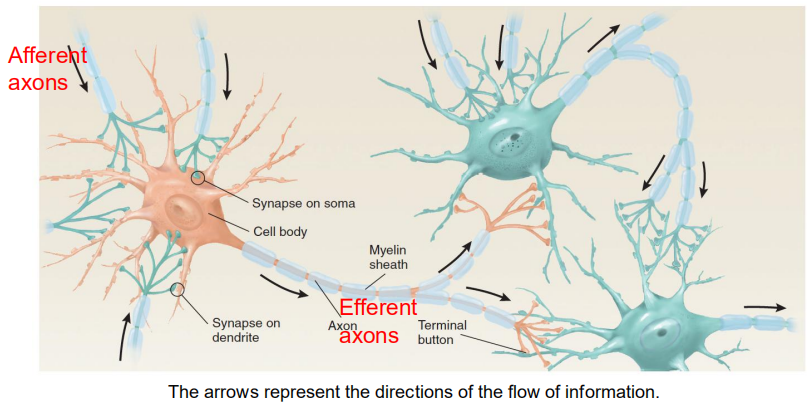
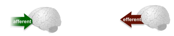
Identify the afferent axon(传入轴突) and efferent axons(传出轴突): Identify the upstream and downstream
Methods:
Anterograde labeling method 顺行标记法
PHA-L
A protein derived from kidney beans and used as an anterograde tracer; taken up by dendrites and cell bodies and carried to the ends of the axons
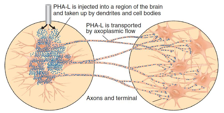
(The diagram illustrates the use of PHA-L to trace efferent axons.)
Retrograde labeling method 逆行标记法
- Fluorogold
- A dye that serves as a retrograde label; taken up by terminal buttons and carried back to the cell bodies.
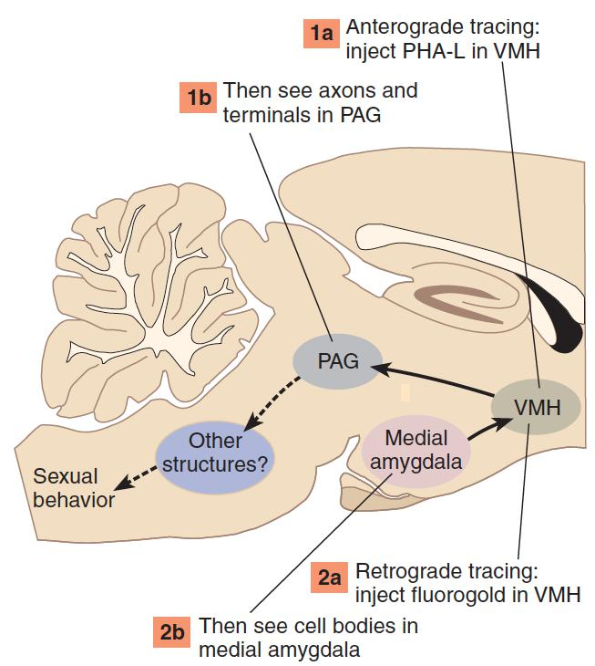
- Fluorogold
Study the structure of living human brain
- Computerized Tomography (CT) 计算机断层扫描
- The use of a device that employs a computer to analyze data obtained by a scanning beam of X-rays to produce a two-dimensional picture of a “slice” through the body.
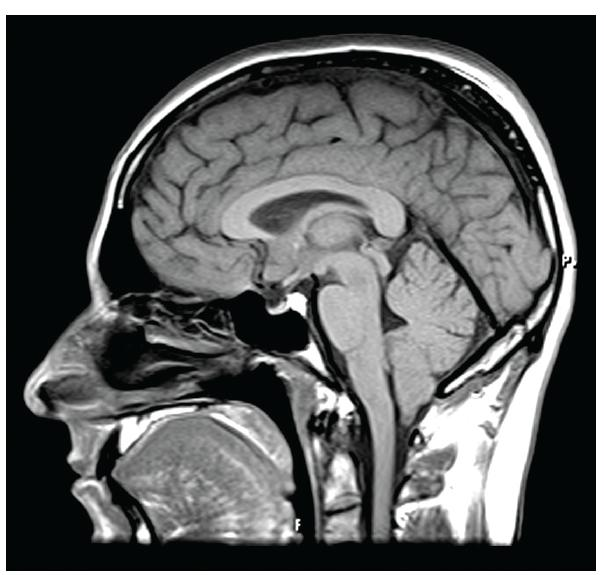
- Magnetic resonance imaging (MRI) 磁共振成像
- A technique whereby the interior of the body can be accurately imaged; involves the interaction between radio waves and a strong magnetic field
- Diffusion tensor imaging (DTI) 扩散张量成像
- An imaging method that uses a modified MRI scanner to reveal bundles of myelinated axons in the living human brain
1. Computerized Tomography (CT)
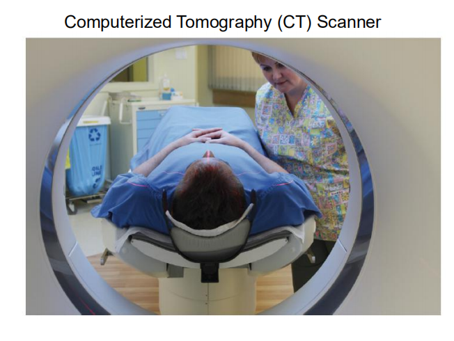
The patient had a lesion in the right occipital-parietal area (scan 5). The lesion appears white because it was accompanied by bleeding; blood absorbs more radiation than does the surrounding brain tissue. Rostral is up, caudal is down; left and right are reversed. Scan 1 shows a section through the eyes and the base of the brain

2. Magnetic Resonance Imaging (MRI)
3. Diffusion Tensor Imaging (DTI)

A sagittal view of some of the axons that project from the thalamus to the cerebral cortex in the human brain is revealed by diffusion tensor imaging.
Recording Neural Activity
Axons produce action potentials, and terminal buttons elicit postsynaptic potentials in the membrane of the cells with which they form synapses.
- These electrical events can be recorded, and changes in the electrical activity of a particular region can be used to determine whether that region plays a role in various behaviors.
- Electrophysiology
- Voltage imaging / Calcium imaging
1. Techniques
- Whole-Cell Patch Clamp
- Current Clamp
- Extracellular Recording
Patch Clamp
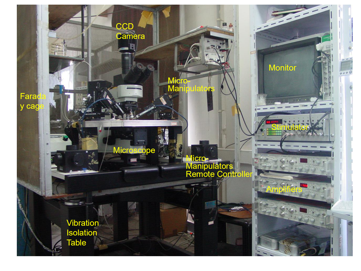

Extracelluar Recording

Recordings can be made chronically, over an extended period of time after the animal recovers from surgery, or acutely, for a relatively short period of time during which the animal is kept anesthetized
If we want to record the activity of a particular region of the brain of an animal that is awake and free to move about, we would implant the electrodes by means of stereotaxic surgery
Then, after recovery from surgery, the animal can be “plugged in” to the recording system. Laboratory animals pay no heed to the electrical sockets on their skulls and behave quite normally

- This schematic shows a permanently attached set of electrodes, with a connecting socket cemented to the skull.
Recording the Brain’s Metabolic and Synapic Activity
Electrical signals are not the only signs of neural activity. If the neural activity of a particular region of the brain increases, the metabolic rate of this region increases, too, largely as a result of increased operation of transporters in the membrane of the cells. This increased metabolic rate can be measured
The experimenter injects radioactive 2-deoxyglucose (2-DG) into the animal’s bloodstream. Because this chemical resembles glucose (the principal food for the brain), it is taken into cells. Thus, the most active cells, which use glucose at the highest rate, will take up the highest concentrations of radioactive 2-DG. But unlike normal glucose, 2-DG cannot be metabolized, so it stays in the cell. The experimenter then kills the animal, removes the brain, slices it, and prepares it for autoradiography
2-deoxyglucose (2-DG)
- A sugar that enters cells along with glucose but is not metabolized
Autoradiography
- A procedure that locates radioactive substances in a slice of tissue; the radiation exposes a photographic emulsion or a piece of film that covers the tissue.
1. 2-DG Autoradiography

- This 2-DG autoradiogram of a rat brain (frontal section, dorsal is at top) shows especially high regions of activity in the pair of nuclei in the hypothalamus, at the base of the brain.
2. Fos
Another method of identifying active regions of the brain capitalizes on the fact that when neurons are activated (for example, by the terminal buttons that form synapses with them), particular genes in the nucleus called immediate early genes are turned on and particular proteins are produced. These proteins then bind with the chromosomes in the nucleus. The presence of these proteins indicates that the neuron has just been activated.
- One of the nuclear proteins produced during neural activation is called Fos .
Fos
- A protein produced in the nucleus of a neuron in response to synaptic stimulation.
- The photomicrograph shows a frontal section of the brain of a female rat, taken through the medial amygdala. The dark spots indicate the presence of Fos protein, localized by means of immunocytochemistry. The synthesis of Fos protein was stimulated by permitting the animal to engage in copulatory behavior.
3. Functional imaging
Positron emission tomography (PET) 正电子发射断层扫描
A functional imaging method that reveals the localization of a radioactive tracer in a living brain. This imaging test that helps reveal how your tissues and organs are functioning and active.
- First, the patient receives an injection of radioactive 2-DG. (The chemical soon breaks down and leaves the cells. The dose given to humans is harmless.)
- The person’s head is placed in a machine similar to a CT scanner.
- When the radioactive molecules of 2-DG decay, they emit subatomic particles called positrons, which meet nearby electrons.
PET Scans:

- In the top row of these human brain scans are three horizontal scans from a person at rest. The bottom row shows three scans from the same person while he was clenching and unclenching his right fist. The scans show increased uptake of radioactive 2-deoxyglucose in regions of the brain that are devoted to the control of movement, which indicates increased metabolic rate in these areas. Different computergenerated colors indicate different rates of uptake of 2-DG, as shown in the scale at the bottom.
Function Magnetic resonance imaging (fMRI)
Function Magnetic resonance imaging (fMRI) : is a technique for measuring brain activity. It works by detecting the changes in blood oxygenation and flow that occur in response to neural activity when a brain area is more active it consumes more oxygen and to meet this increased demand blood flow increases to the active area.
The formal name of this type of imaging is BOLD—blood oxygen level-dependent signal. Functional MRI scans have a higher resolution than PET scans, and they can be acquired much faster. Thus, they reveal more detailed information about the activity of particular brain regions
Functional MRI Scans:
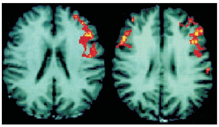
These scans of human brains show localized average increases in neural activity of males (left) and females (right) while they were judging whether pairs of written words rhymed
Stimulating Neural Activity
- Electrical stimulation
- Electrical stimulation simply involves passing an electrical current through a wire inserted into the brain.
- Chemical stimulation
- Chemical stimulation is usually accomplished by injecting a small amount of an excitatory amino acid, such as kainic acid or glutamic acid, into the brain.
1. Deep Brain Stimulation

- Deep brain stimulation involves implanting electrodes within certain areas of your brain. These electrodes produce electrical impulses that regulate abnormal impulses. Or the electrical impulses can affect certain cells and chemicals within the brain.
- Deep brain stimulation is an established treatment for people with movement disorders, such as essential tremor, Parkinson’s disease and dystonia, and psychiatric conditions, such as obsessive- compulsive disorder.
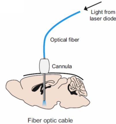
2. Optogenetic Method
Optogenetic methods can be used to stimulate or inhibit particular types of neurons in particular brain regions (Boyden et al., 2005; Zhang et al., 2007; Baker, 2011)
Photosensitive proteins have evolved in many organisms—even single-celled organisms such as algae and bacteria. Researchers have discovered that when blue light strikes one of these proteins, (ChR2), the channel opens, and the rush of positively charged sodium and calcium ions depolarizes the membrane, causing excitation
The use of a genetically modified virus to insert light sensitive ion channels into the membrane of particular neurons in the brain; can depolarize or hyperpolarize the neurons when light of the appropriate wavelength is applied.
3. Pharmacogenetic (chemogenetic) activation
Use genetic technique to activate the activity of specific neuron populations using DREDD(designer receptors exclusively activated by designer drugs)
4. Transcranial magnetic stimulation (TMS)
Stimulation of the cerebral cortex by means of magnetic fields produced by passing pulses of electricity through a coil of wire placed next to the skull; interferes with the functions of the brain region that is stimulated.
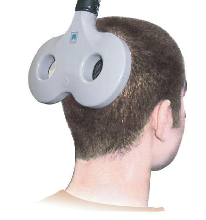
- Pulses of electricity through the coil produce a magnetic field that stimulates a region of the cerebral cortex under the crossing point in the middle of the figure
Genetic Methods
All behavior is determined by interactions between an individual’s brain and his or her environment
Many behavioral characteristics—such as talents, personality variables and mental disorders—seem to run in families
This fact suggests that genetic factors may play a role in the development of physiological differences that are ultimately responsible for these characteristics
The methods involves:
- Twin Studies
- Adoption Studies
- Genomic Studies
- Targeted Mutations
- Antisense Oligonucleotides
1. Twin Study
A powerful method for estimating the influence of heredity on a particular trait is to compare the concordance rate for this trait in pairs of monozygotic and dizygotic twins.
- Monozygotic twins (identical twins) have identical genotypes—that is, their chromosomes, and the genes they contain, are identical.
- In contrast, the genetic similarity between dizygotic twins (fraternal twins) is, on the average, 50 percent.
Investigators study records to identify pairs of twins in which at least one member has the trait—for example, a diagnosis of a particular mental disorder.
- If both twins have been diagnosed with this disorder, they are said to be concordant.
- If only one has received this diagnosis, the twins are said to be discordant.
2. Adoption Studies
Adoption studies require that the investigator know the identity of the parents of the people being studied and be able to measure the behavioral trait in the biological and adoptive parents.
If the people being studied strongly resemble their biological parents, we conclude that the trait is probably influenced by genetic factors.
- To be certain, we will have to rule out possible differences in the prenatal environment of the adopted children.
Genome
- The complete set of genes that compose the DNA of a particular species.
Allele
- The nature of the particular sequence of base pairs of DNA that constitutes a gene; for examples, the genes that code for blue or brown iris pigment are different alleles of a particular gene
Targeted mutation
- A mutated gene (also called a “knockout gene”) produced in the laboratory and inserted into the chromosomes of mice; fails to produce a functional protein.
Antisense oligonucleotide
- A modified strand of RNA or DNA that binds with a specific molecule of messenger RNA and prevents it from producing its particular protein.