L8 Kidney
一、Kidney and Excretion
Structure of mammalian kidney
1. What are kidneys?
Animals have various types of kidneys, but their structures have three features in common:
- Tubular elements that discharge directly or indirectly to the outside
- Produce or eliminate aqueous solutions derived from the blood
- They all function in regulating the composition and volume of the blood plasma by means of controlling excretion of solutes and water
Urine – the product of the kidneys is typically a complex solution containing multiple inorganic and organic solutes.
- Primary urine (原发性尿)
- Definite urine (定尿) that is eliminated

Primary urine is introduced into the kidney tubules by ultrafiltration
ultrafiltrationa filtration process for water and a subset of solutes that are able to stream through the nephron wall (肾单位).
The blood pressure produced by the heart is typically the pressure that drives ultrafiltration
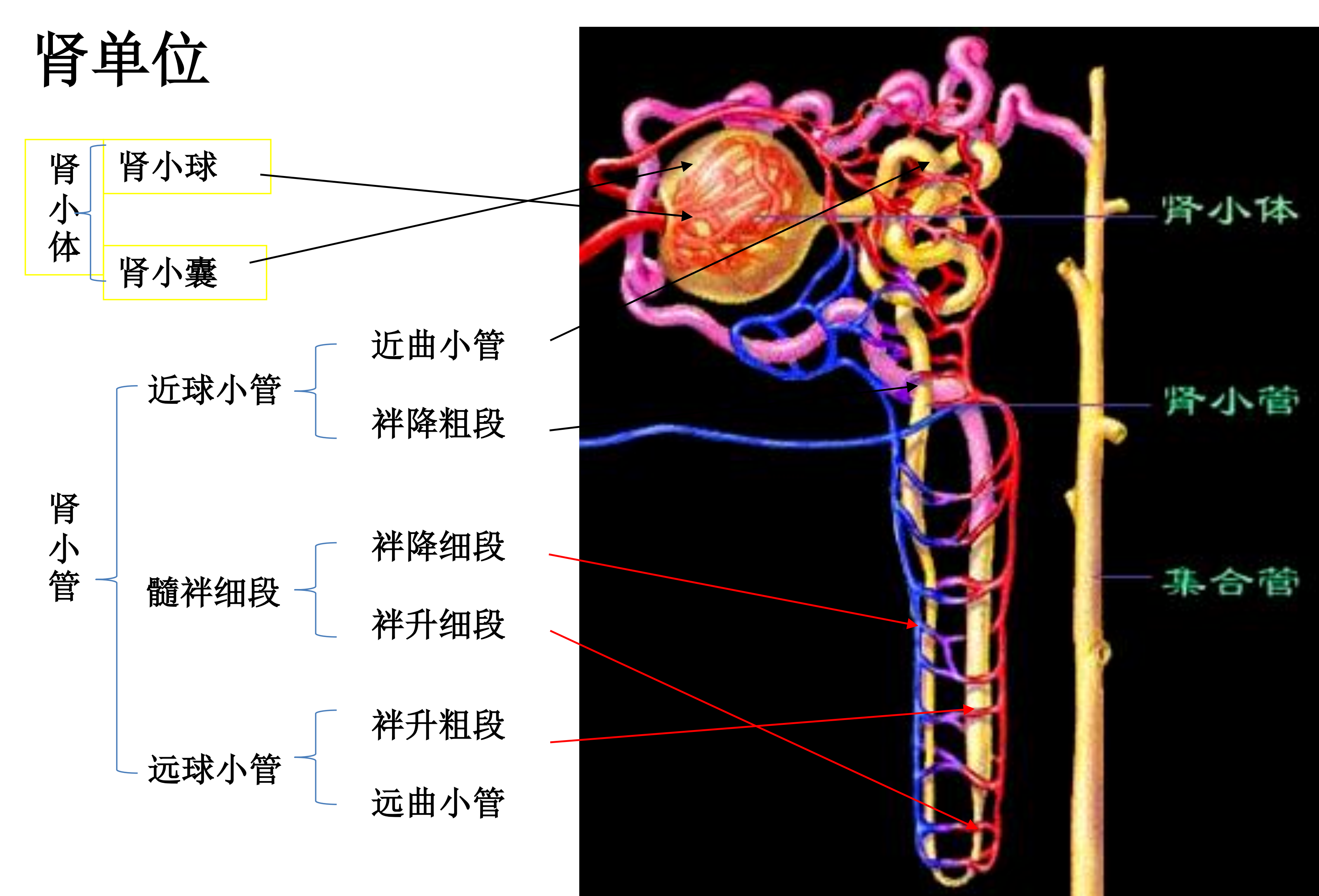
2. Nephron 肾单位
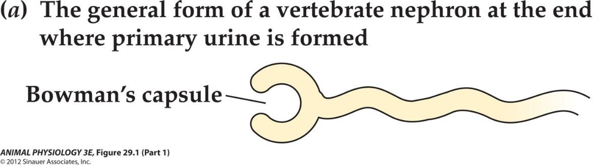
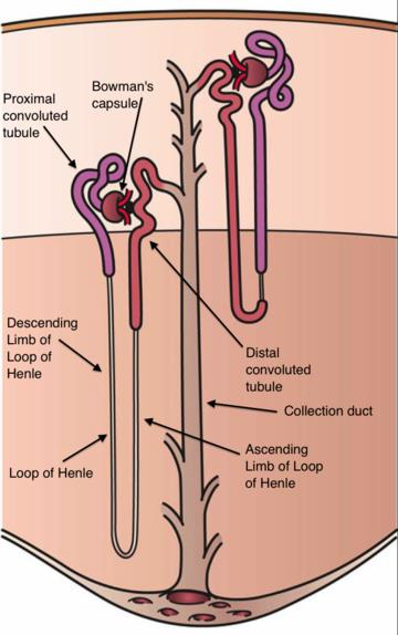
The nephron **(肾单位)**is the microscopic structural and functional unit of the kidney. It is composed of a **renal corpuscle(肾小体, ** and a renal tubule(肾小管, .
The renal corpuscle consists of a tuft of capillaries called a **glomerulus(肾小球, ** and an encompassing Bowman’s capsule. The renal tubule extends from the capsule. Nephron’s wall consists of only a single layer of epithelial cells
3. Bowman’s capsule 肾小球囊
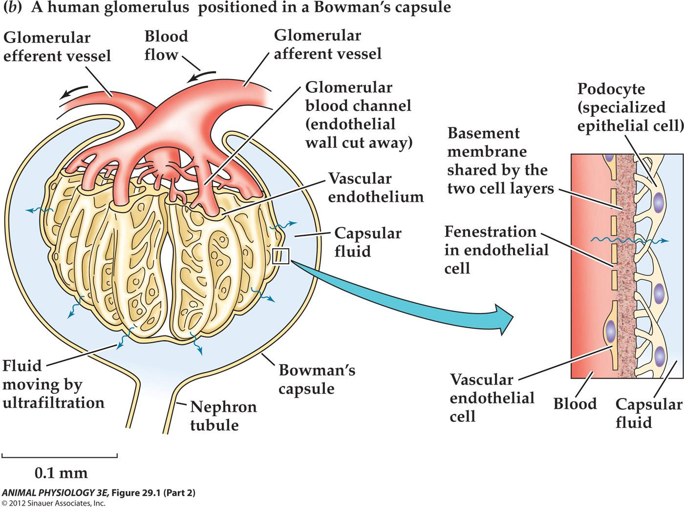
Capsular fluid is the primary urine
4. glomerulus 肾小球
Glomerulus – Cluster of blood capillaries
Some terms:
- glomerulus 肾小球
- Ultrafiltration 超滤
- Capsular fluid 囊液
- Fenestration 开窗
- Colloid osmotic pressure 胶体渗透压
- Capsular fluid hydrostatic pressure 囊液静水压力
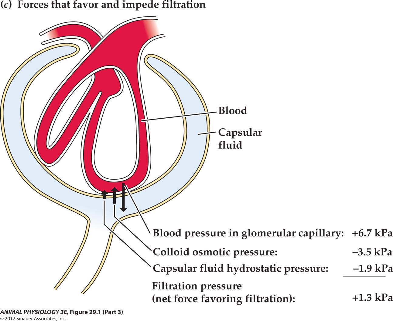
肾小球有效过滤压 = 毛细血管血压 - (血浆胶体渗透压 + 囊内压)
Basic Mechanisms of Kidney Function
1. Forces that favor and impede (阻碍) filtration
- Podocytes 足细胞
- Slit diaphragm
Small organic ions and inorganic ions move freely with filtered fluid as it passes from the blood plasma into the lumen of a Bowman’s capsule.
- 10kD proteins will not be able to pass through.
So the primary urine is devoid of high molecular weight organic solutes such as proteins. Therefore, the osmotic pressure of blood plasma is higher than the osmotic pressure of the capsular fluid – called colloid osmotic pressure of the blood
Water movement between the blood plasma and capsular fluid
TWO processes tend to cause water movement
- Difference in osmotic pressure causing the osmosis of water from capsular fluid into the blood plasma
- Difference in hydrostatic pressure tends to cause osmosis of water from the capsular fluid into the blood (Blood pressure)
Net flow of fluid into the capsular lumen will occur only if the hydrostatic pressure is greater than the difference in osmotic pressure – the net force favors filtration – filtration pressure
Hydrostatic pressure is the pressure that is exerted by a fluid at equilibrium at a given point within the fluid, due to the force of gravity. Hydrostatic pressure increases in proportion to depth measured from the surface because of the increasing weight of fluid exerting downward force from above
Filtration Rate
Filtration rate: the rate of primary urine formation
- By all of an animals kidney tubules taken together!
- In vertebrates, it is called glomerular filtration rate (GFR)
For an adult human, a GFR is about 120 ml/min At this rate the equivalent of all the plasma water in a person’s body is filtered about every 30 min!
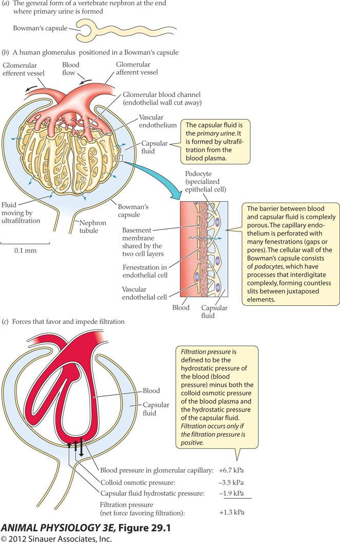
- The capsular fluid is the primary urine. It is formed by ultrafiltration from the blood plasma.
- The barrier between blood and capsular fluid is complexly porous. The capillary endothelium is perforated with many fenestrations(gaps or pores). The cellular wall of the Bowman’s capsule consists of podocytes, which have processes that interdigitate complexly, forming countless slits between juxtaposed elements
- Filtration pressure is defined to be the hydrostatic pressure of the blood(blood pressure) minus both the colloid osmotic pressure of the blood plasma and the hydrostatic pressure of the capsular fluid Filtration occurs only if the filtration pressure is positive
Urine Formation in Mammals
1. Formation of primary urine by active solute secretion
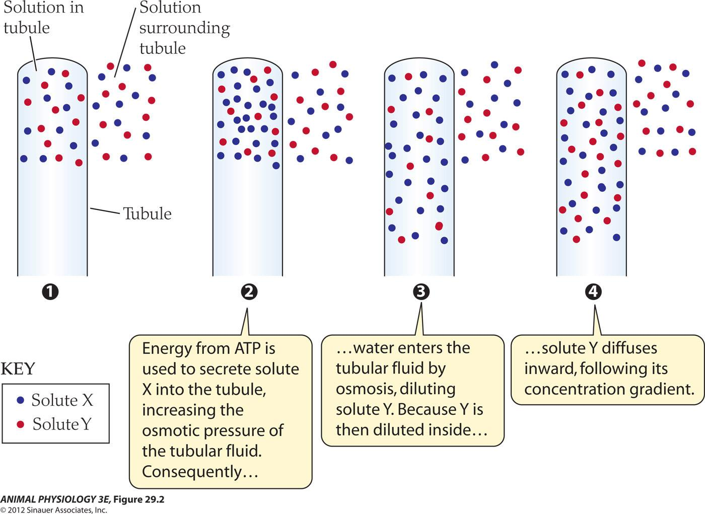
Primary urine formation: 浓缩尿液:
- Energy from ATP is used to secrete solute X into the tubule, increasing the osmotic pressure of the tubular fluid
- Consequently, water enters the tubular fluid by osmosis, diluting solute Y in tubule
- Because Y is then diluted inside, solute Y diffuses inward, following its concentration gradient
2. Regulatory processes in kidney
Most of the water in the primary urine is usually reabsorbed and returned to the blood plasma.
Hyperosmotic urine The presence of loop of Henle(Hen-lee) and their parallel arrangement are the basis to produce urine that is osmotically concentrated than blood.
Frogs, snakes turtles – lack of these anatomical features and can not produce hyperosmotic urine.
Ability to concentrate urine is one of the most important adaptions for life on land
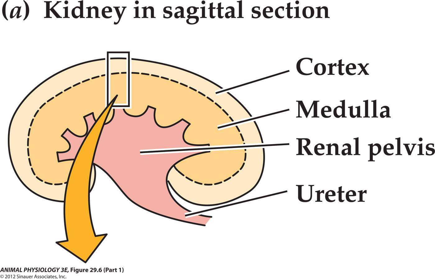
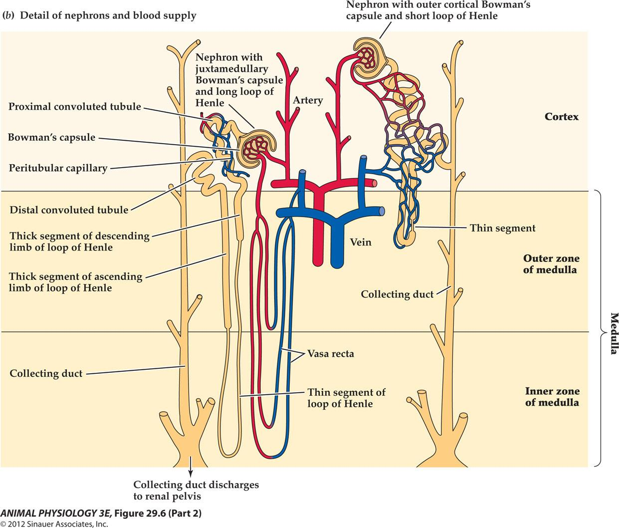
肾的功能解剖和肾血流量示意图:
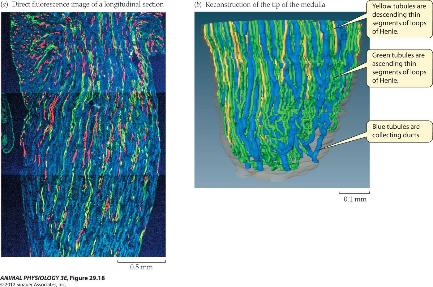
3. The role of loops of Henle in concentrating the urine
Freshwater species lack of Henle and have only short loops, no inner medulla and have very limited capacity to concentrate their urine
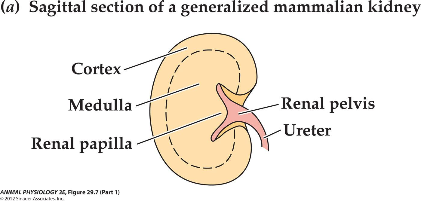
Renal papilla – not all animals have apparent renal papilla, which consist of mainly The long loops of Henle.
The medullary thickness, relative medullary thickness ( a ratio of medulla thickness With the absolute size of kidney).
The papilla is composed in major part of the long loops of Henle
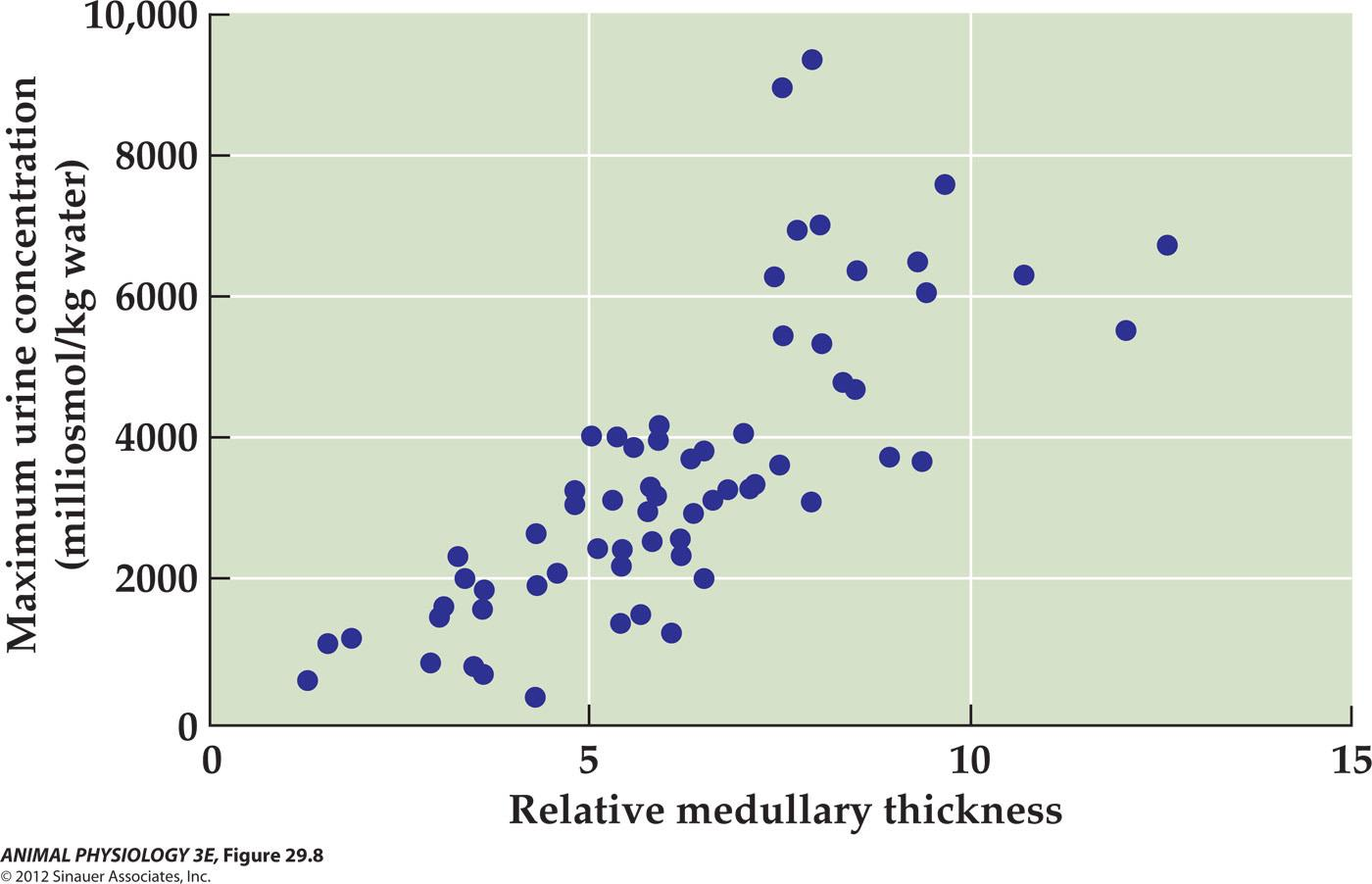
Maximum urine concentration correlates with the relative thickness of the medulla
a plot of 68 mammalian species:
4. 尿液浓缩的机理—逆流学说
Countercurrent multiplication is the key to producing concentrated urine
- Urea
- Nonurea solutes (Na+, K+, and SO42-)
- The concentration of urine: countercurrent multiplication is the key to producing concentrated urine
Generation of the single effect in the loop of Henlee
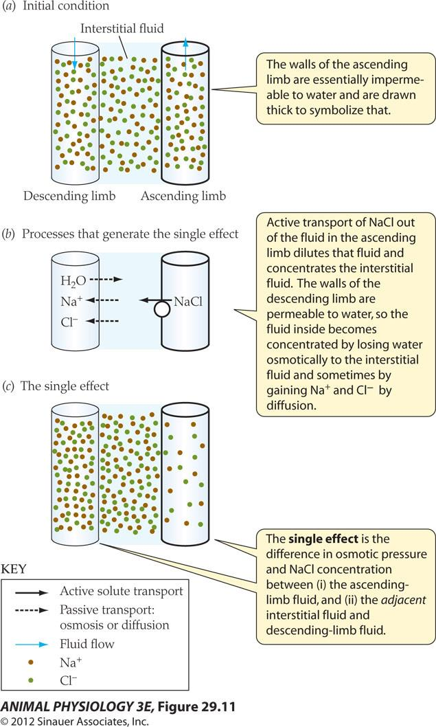
- The walls of the ascending limb are essentially impermeable to water and are drawn thick to symbolize that
- Active transport of Nacl out of the fluid in the ascending limb dilutes that fluid and concentrates the interstitial fluid. The walls of the descending limb are permeable to water, so the fluid inside becomes concentrated by losing water osmotically to the interstitial fluid and sometimes by gaining Na”and Cr-by diffusion
- The single effect is the difference in osmotic pressure and NACI concentration between(i)the ascending limb fluid, and (ii)the adjacent interstitial fluid and descending-limb fluid
A single effect based on active NaCl transport
- The walls of the ascending limb are essentially impermeable to water.
- The active transport of NaCl out of the Fluid in the ascending limb.
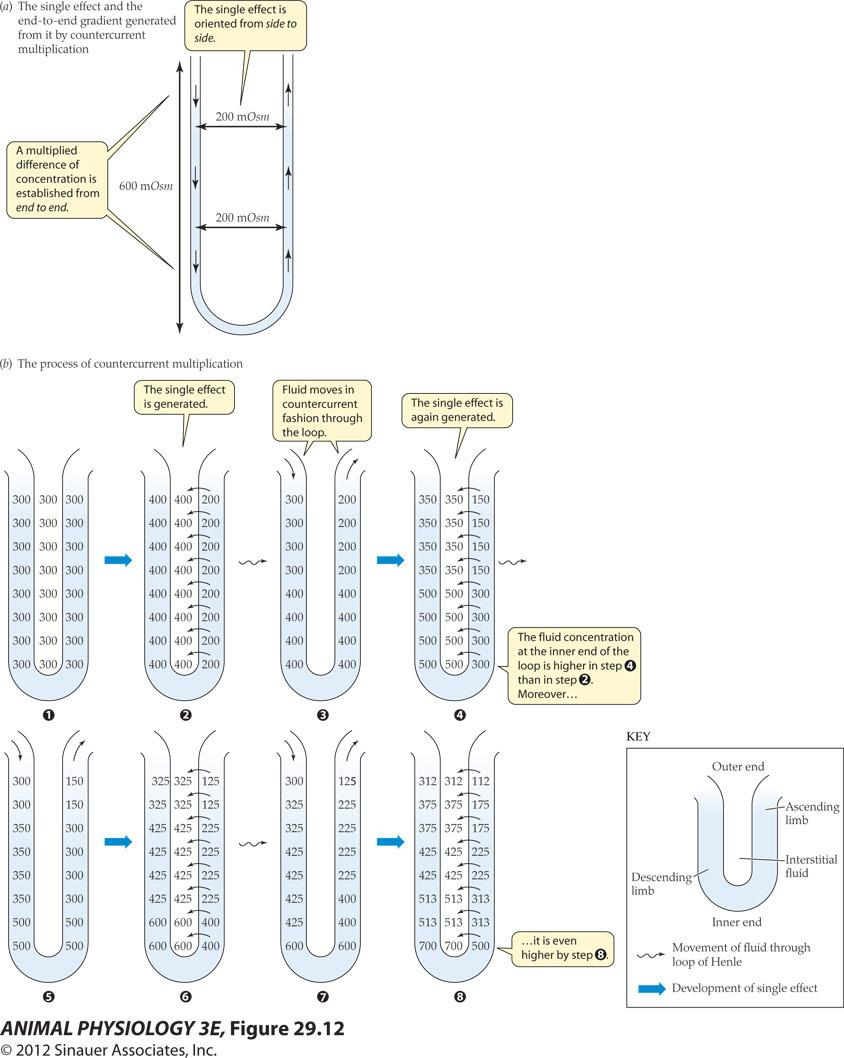
A single effect based on active NaCl transport
- Ascending thick segment of a loop of Henle actively transport NaCl from the tubular fluid inside the loop into the adjacent medullary interstitial fluid
Osmotic pressures attributable to nonurea solutes in the nephrons and collecting ducts during antidiuresis and diuresis
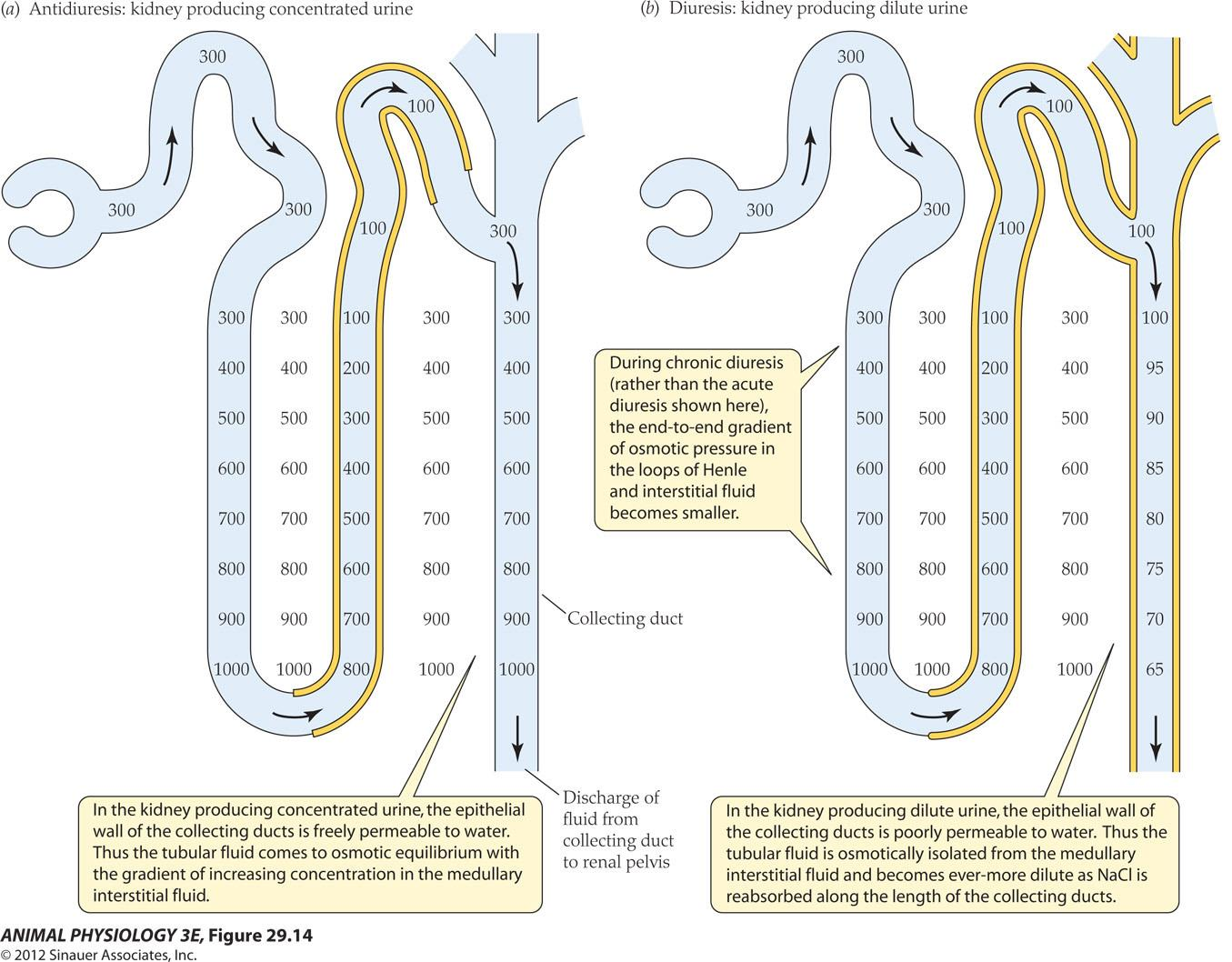
In the kidney producing concentrated urine, the epithelial wall of the collecting ducts is freely permeable to water. Thus the tubular fluid comes to osmotic equilibrium with the gradient of increasing concentration in the medullary interstitial fluid
During chronic diuresis (rather than the acute diuresis shown here) the end-to-end gradient of osmotic pressure in the loops of Henle and interstitial fluid becomes smaller
In the kidney producing dilute urine, the epithelial wall of the collecting ducts is poorly permeable to water. Thus the tubular fluid is osmotically isolated from the medullary interstitial fluid and becomes ever-more dilute as Nacl is reabsorbed along the length of the collecting ducts.
Yellow line Symbolize tubules Are poorly permeable To water
Osmotic pressure increases with depth in the medulla
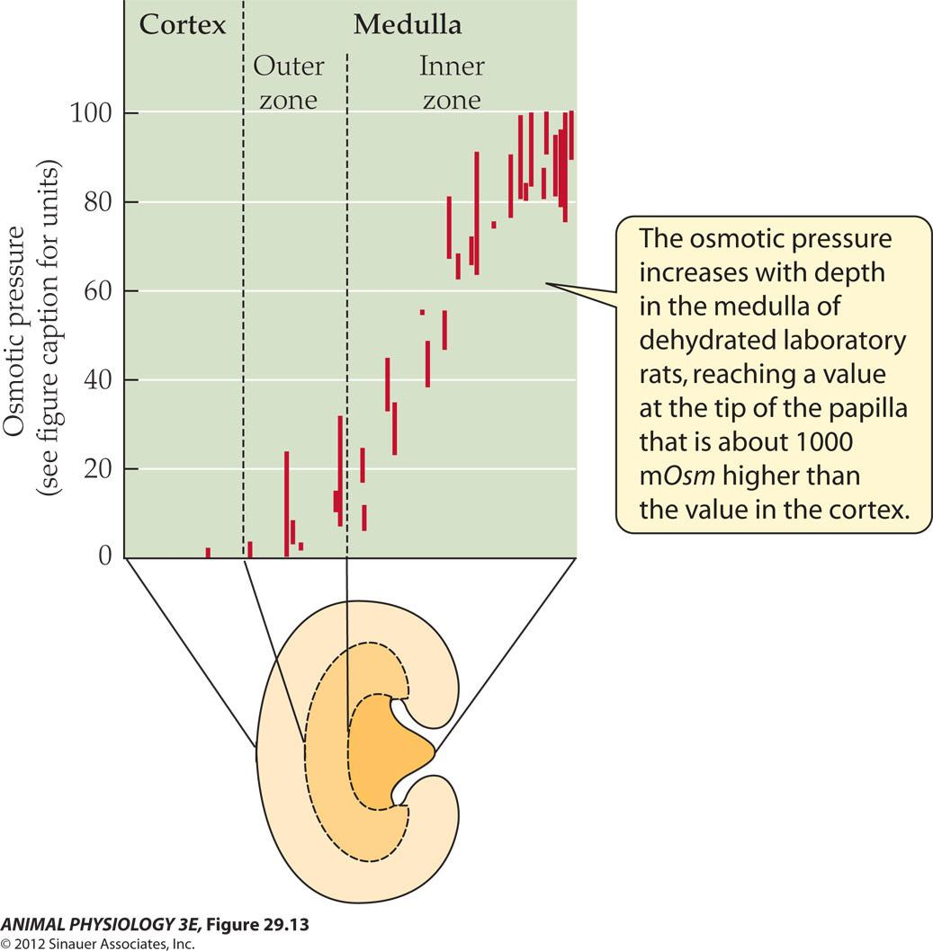
- The osmotic pressure increases with depth in the medulla of dehydrated laboratory rats, reaching a value at the tip of the papilla that is about 1000 mOsm higher than the value in the cortex
5. The regulation of switches between concentration and dilution
A person in antidiuresis can produce urine of 1200 mOsm
A person in diuresis can produce urine as diluted as 50 mOsm
Antidiuretic hormone (ADH,抗利尿激素) is the switch, also called vasopressin. Vasopressin modulates the permeability of the collecting ducts to water.
High blood level of ADH caused the insertion of AQP2 in the apical cell membranes of the collecting duct epithelial cells and increases in epithelial permeability to water.
When blood level ADH decreases, AQP is retrieved from the apical cell membrane.
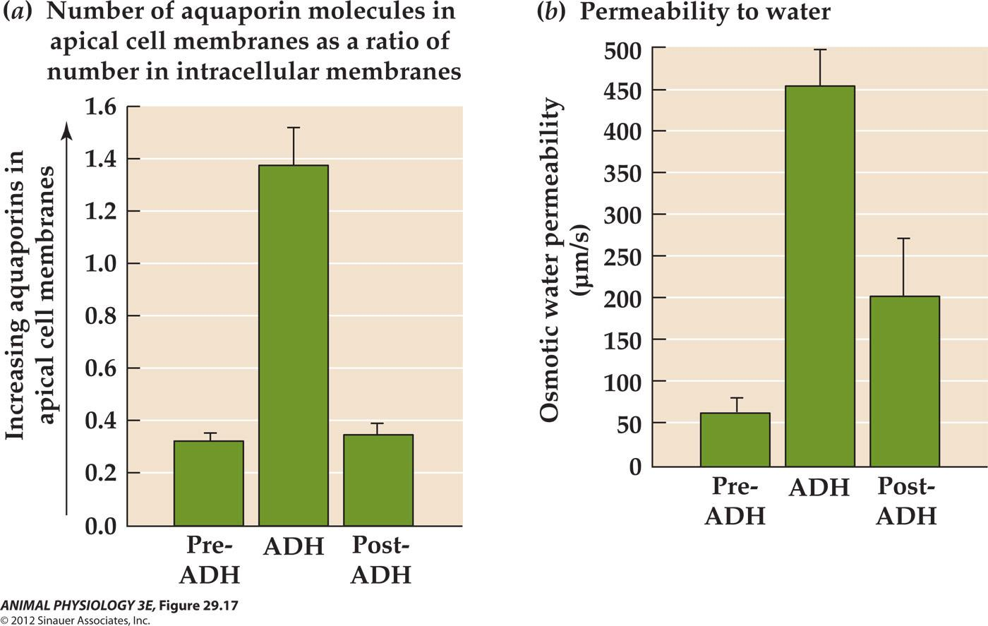
The collecting-duct epithelium: Cellular position of aquaporin-2 (AQP-2) and permeability to water when ADH is present or absent
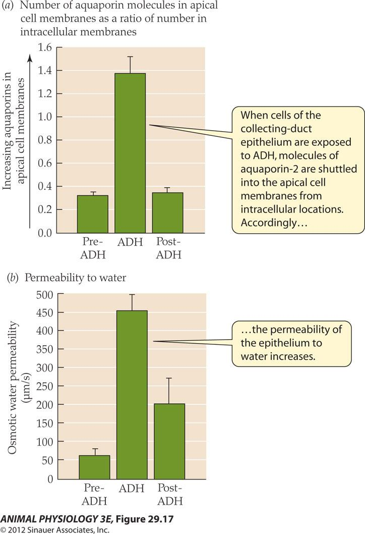
- When cells of the collecting-duct epithelium are exposed to ADH, molecules of aquaporin-2 are shuttled into the apical cell membranes from intracellular locations Accordingly, the permeability of the epithelium to water increases.
The high water permeability permits water to leave the collecting ducts by following the osmotic gradient between the collecting duct fluid and medullary interstitial fluid.
The osmotic exit of water accounts for both the low volume and high concentration of the urine produced during antidiuresis
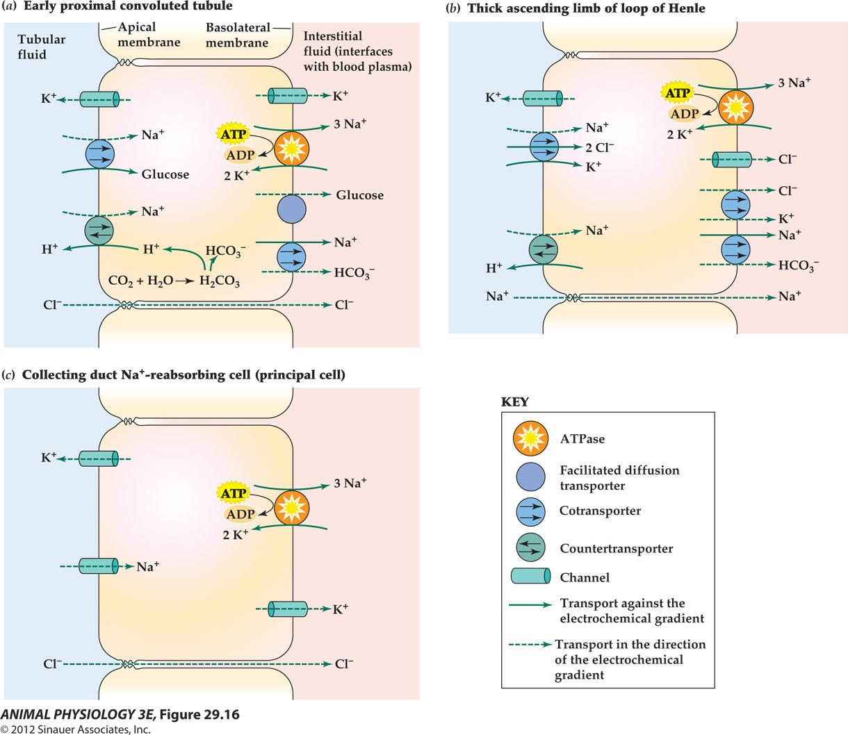
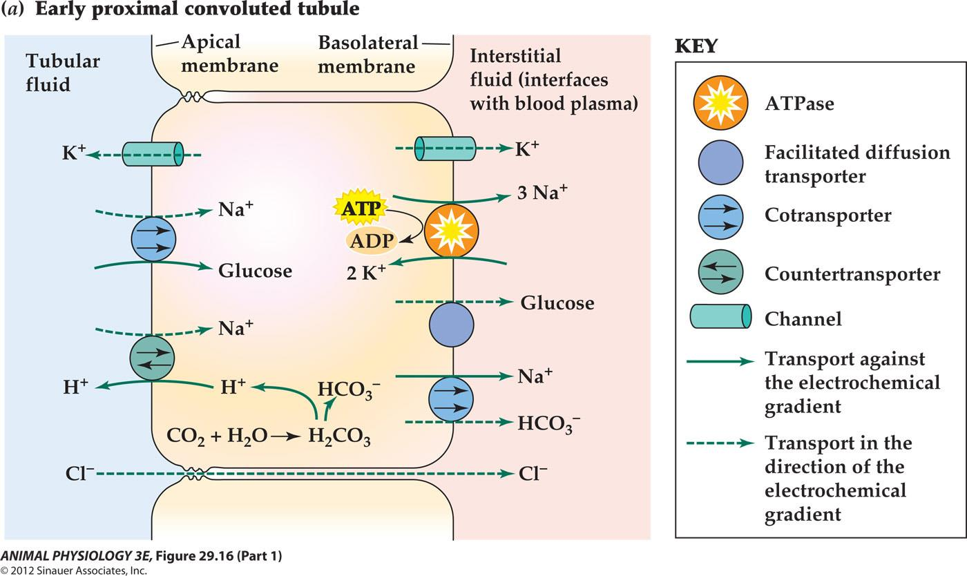
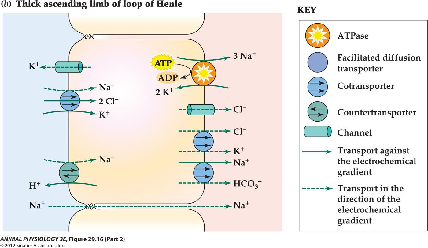
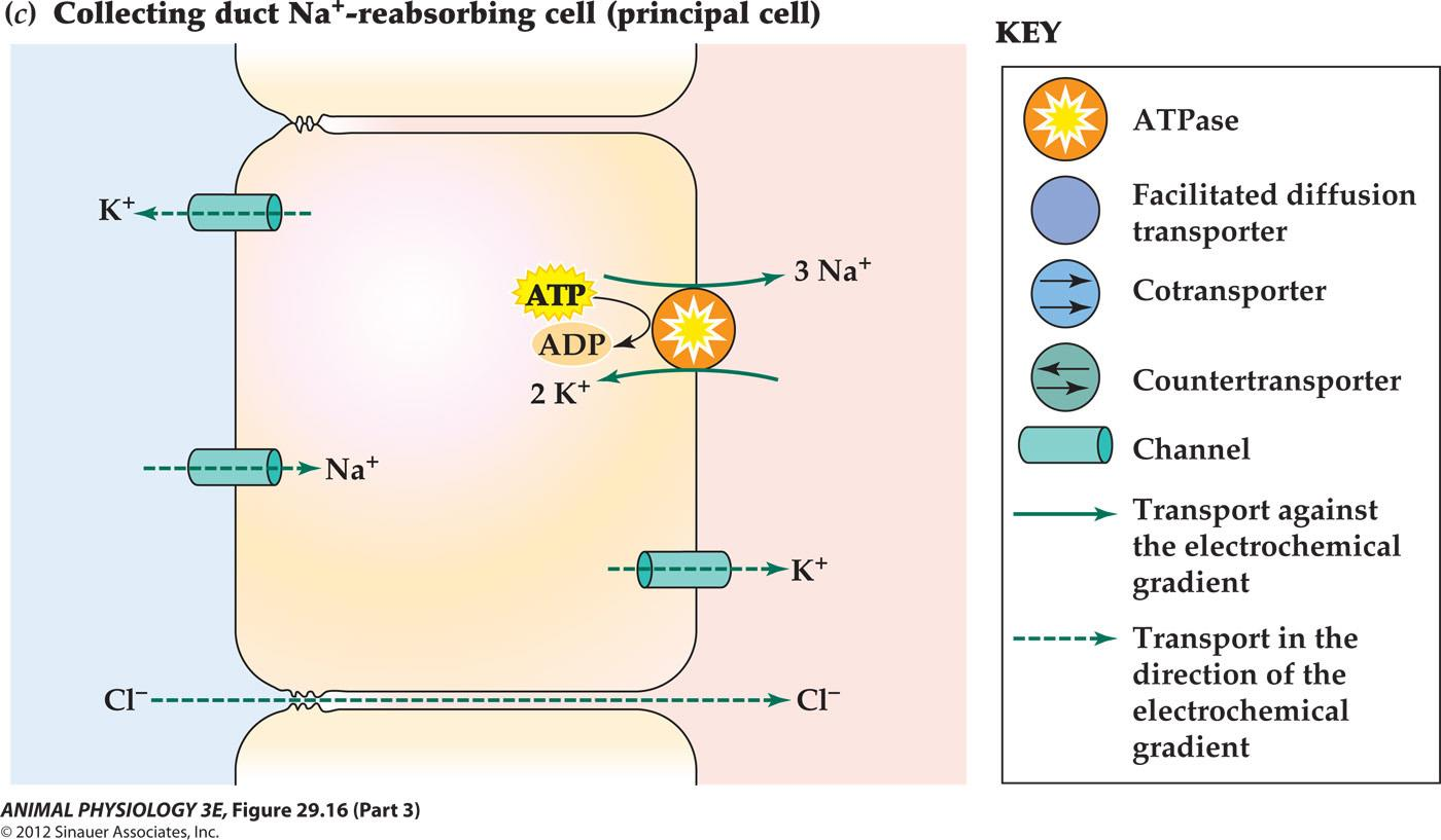
Major molecular mechanisms of NaCl reabsorption and associated processes in three parts of the mammalian kidney tubule
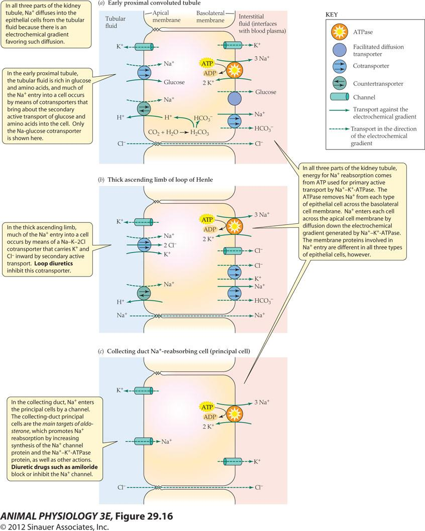
- In all three parts of the kidney tubule. Na+ diffuses into the epithelial cells from the tubular fluid because there is an electrochemical gradient favoring such diffusion
- In the early proximal tubule. the tubular fluid is rich in glucose and amino acids, and much of the Na+ entry into a cell occurs by means of cotransporters that bring about the secondary active transport of glucose and amino acids into the cell. Only the Na-glucose cotransport is shown here
- In the thick ascending limb. much of the Na+ entry into a cell occurs by means of a Na-K-2Cl cotransporter that carries K+ and Cl- inward by secondary active transport. Loop diuretics inhibit this cotransporter
- In the collecting duct, Na+ enters the principal cells by a channel The collecting-duct principal cells are the main targets of aldosterone, which promotes Na reabsorption by increasing synthesis of the Na+ channel protein and the Na+-K+-ATPase protein, as well as other actions. Diuretic drugs such as amiloride block or inhibit the Na+ channel
- In all three parts of the kidney tubule. energy for Na+ reabsorption comes from ATP used for primary active transport by Na+-K+-ATPase. The ATPase removes Na+ from each type of epithelial cell across the basolateral cell membrane Na+ enters each cell across the apical cell membrane by diffusion down the electrochemical gradient generated by Na-K-ATPase The membrane proteins involved in Na+ entry are different in all three types of epithelial cells, however.

Major molecular mechanisms of NaCl reabsorption and associated processes in three parts of the mammalian kidney tubule
6. Control of water and salt balance in terrestrial animals.
Antidiuresis (抗利尿)hormones and diuresis (利尿) hormones
Vertebrates only produce antidiuretic hormones
- ADH produced by neurohypophysis(神经垂体)
- mineralocorticoids (盐皮质激素) (aldosterone, 醇固酮)
- Natriuretic hormones (利钠激素)
The principal agent of control of switches between antidiuresis and diuresis is antidiuretic hormone (ADH). The ADH of most mammals is arginine vasopressin; therefore, ADH is often called vasopressin in books on mammalian physiology and medicine.
- The effect of ADH on the permeability of the collecting duct epithelium is mediated by a specific molecular form of aquaporin, AQP-2, that is inserted into and retrieved from the apical cell membranes of the collecting-duct epithelial cells; Figure 16.16 shows this process in detail.
When ADH levels fall, the aquaporin molecules are retrieved from the apical cell membranes and epithelial permeability to water decreases.
The collecting-duct epithelium: Cellular position of aquaporin-2 (AQP-2) and permeability to water when ADH is present or absent
7. An antidiuretic hormone functions to conserve water
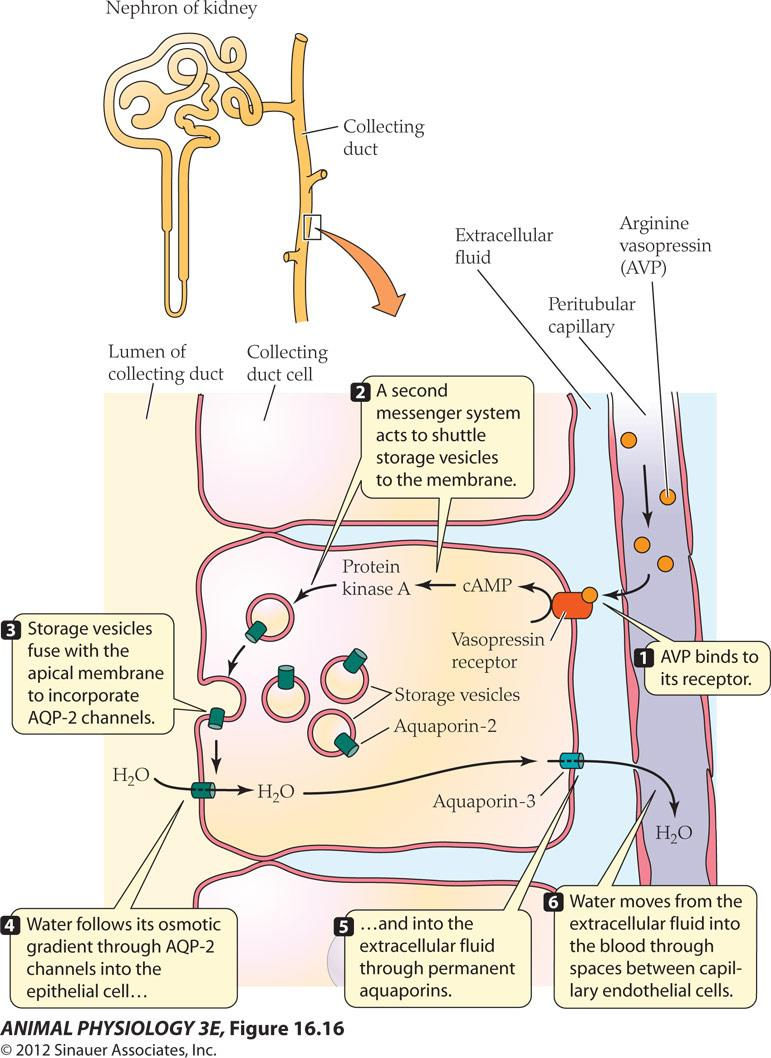
- AVP binds to its receptor.
- A second messenger system acts to shuttle storage vesicles to the membrane
- Storage vesicles fuse with the apical membrane to incorporate AQP-2 channels
- Water follows its osmotic gradient through AQP-2 channels into the epithelial cells
- and into the extracellular fluid through permanent aquaporins.
- Water moves from the extracellular fluid into the blood through spaces between capillary endothelial cells
FIGURE 16.16: An antidiuretic hormone functions to conserve water Arginine vasopressin (AVP) stimulates the incorporation of aquaporin 2 (AQP-2) into the apical membranes of epithelial cells in the collecting duct of the nephron, resulting in the return of water to the extracellular fluid. The water undergoes osmosis into nearby capillaries and is carried out of the kidney in the bloodstream
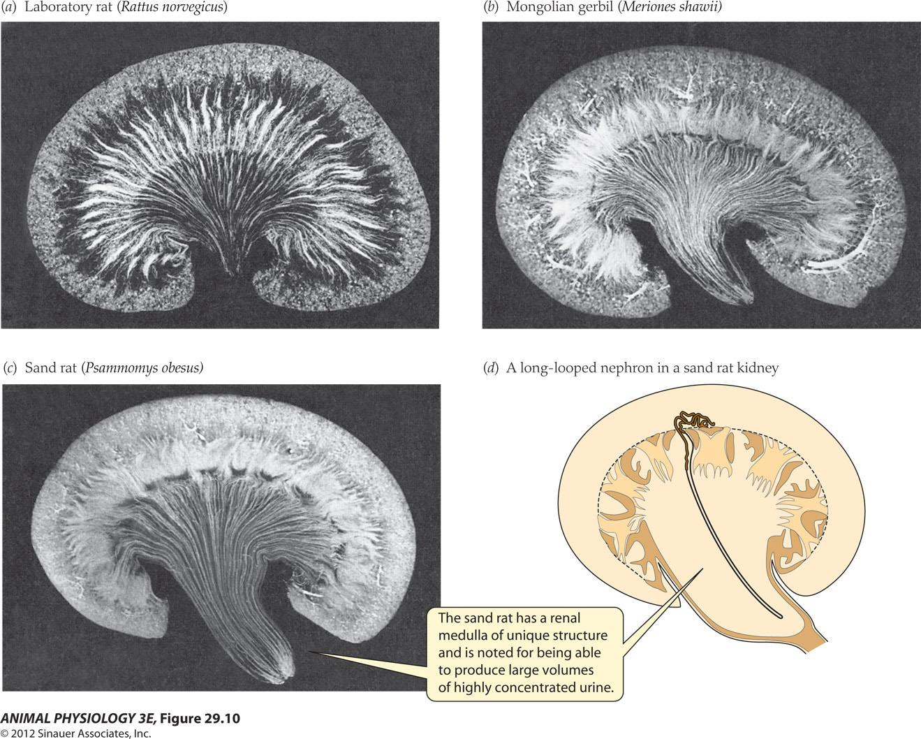
二、Evolutionary development of the renal papilla in mammals native to different habitats
Studies of the fine structure of the papilla of the medulla in young laboratory rats
- Blue = collecting duct
- Red = descending thin segment of the loop of Henle
- Green = descending vasa recta
Kidney structure visualized by injection of the microvasculature
Concentration of urea:
- Urea is present at high concentrations in the medullary interstitial fluid
- The walls of the collecting ducts in the inner medulla permit free diffusion of urea between the urine inside the ducts and the inner – medullary interstitial fluids, facilitated by the urea transporter protein
Outline how the orientation of nephrons relative to each other imparts gross structure to the kidneys of mammal
Nitrogen Disposition and Excretion
When ammonia (NH3 ) or the ammonium ion (NH4+) is the principal nitrogenous end product of an animal, the animal is described as ammonotelic.
If urea is the principal nitrogenous end product, an animal is termed ureotelic. If uric acid is the principal end product, an animal is uricotelic.
- Ammonotelism is the primitive state
- Urea is more costly to synthesize but less toxic than ammonias
1. Some nitrogenous compounds excreted by animals
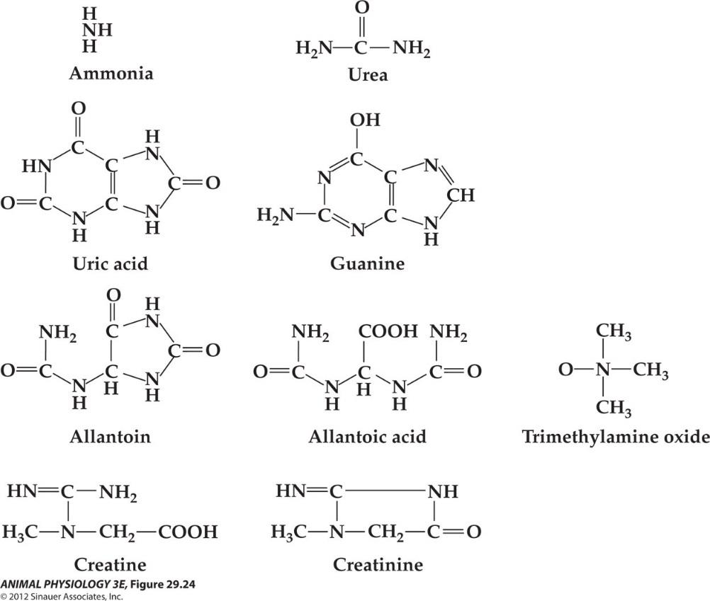
- Some nitrogenous compounds excreted by animals Uric acid and guanine are purines. Allantoin and allantoic acid are poorly soluble breakdown products of uric acid. Trimethylamine oxide and its precursor, trimethylamine, are found in a variety of marine animals but do not occur in freshwater animals; both are highly soluble.
- Creatine and its internal anhydride, creatinine, occur as relatively minor excretory compounds in many vertebrates and some invertebrates. Some animals, mostly invertebrates, also lose significant amounts of amino acids to the environment.
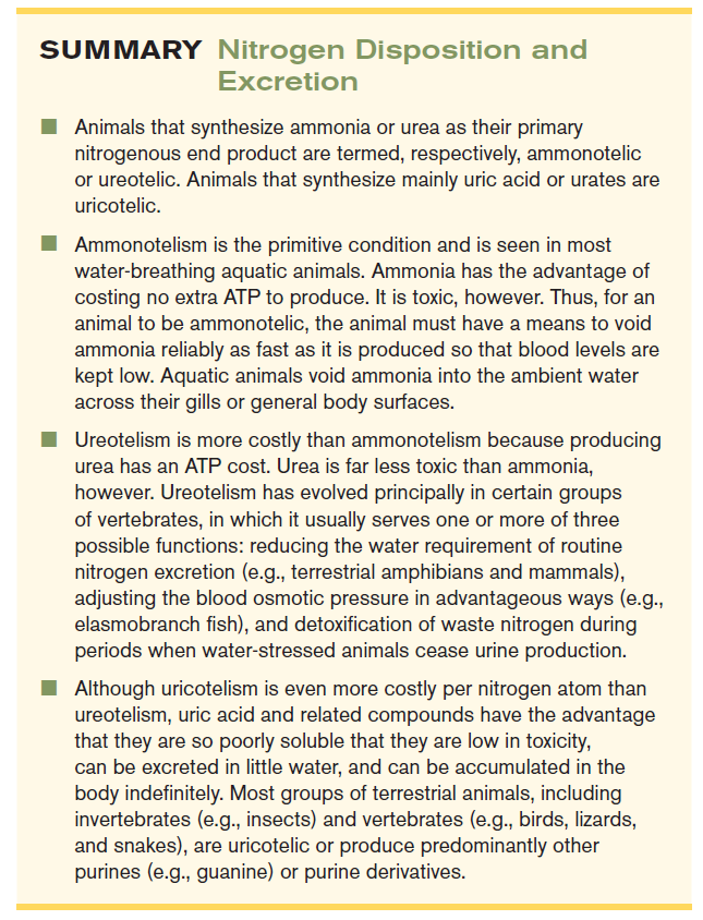
- Animals that synthesize ammonia or urea as their primary nitrogenous end product are termed, respectively, ammonotelic or ureotelic. Animals that synthesize mainly uric acid or urates are uricotelic
- Ammonotelism is the primitive condition and is seen in most water-breathing aquatic animals. Ammonia has the advantage of costing no extra ATP to produce. It is toxic, however. Thus, for an animal to be ammonotelic. the animal must have a means to void ammonia reliably as fast as it is produced so that blood levels are kept low. Aquatic animals void ammonia into the ambient water across their gills or general body surfaces
- Ureotelism is more costly than ammonotelism because producing urea has an ATP cost. Urea is far less toxic than ammonia however Ureotelism has evolved principally in certain groups of vertebrates, in which it usually serves one or more of three possible functions: reducing the water requirement of routine nitrogen excretion(e.g, terrestrial amphibians and mammals) adjusting the blood osmotic pressure in advantageous ways(e.g elasmobranch fish), and detoxification of waste nitrogen during periods when water-stressed animals cease urine production
- Although uricotelism is even more costly per nitrogen atom than ureotelism. uric acid and related compounds have the advantage that they are so poorly soluble that they are low in toxicity, can be excreted in little water and can be accumulated in the body indefinitely. Most groups of terrestrial animals, including invertebrates (e.g. insects)and vertebrates(e.g, birds, lizards, and snakes), are uricotelic or produce predominantly other purines (e.g, guanine)or purine derivatives