L10 Synapses
一、Synaptic strength underlies learning and memory
Synaptic functions
Synaptic transmission
- Action potential – presynaptic signal – is rapid and transient
- Can be excitatory or inhibitory
- Neuromuscular junction as an example
Synaptic plasticity
- The ability to change the synapse functional properties, to make it stronger or weaker which produces long lasting changes of the postsynaptic cells.
A synapse can work both chemically or electrically
There are two kinds of synapses: chemical and electrical;
- both kinds can rapidly change the membrane potential of a postsynaptic cell.
Electrical and chemical synapses
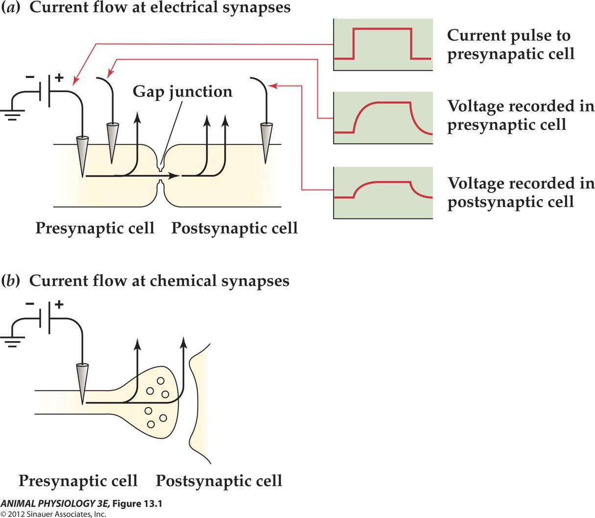
Electrical synapses have low-resistance pathways that allow currents to flow directly between neurons.
Chemical synapses the currents escape between neurons and do not enter the postsynaptic neuron.
- Neurotransmitter are released.
- Most synaptic transmission is chemical, While direct electrical transmission occurs uncommonly
Electrical synapses transmit signals instantaneously
- Signals have no delay
- Can be both directions (not polarized)
- Can be used for synchronization
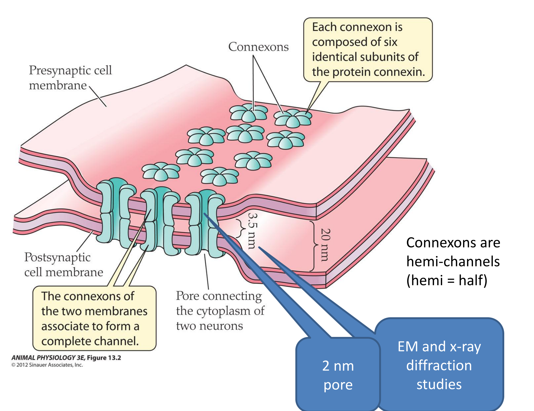
- GAP junction is the major structure for electrical synapse
1. Electrical Synapses
The molecular structure of gap junctions
Electrical synapses in the crayfish escape circuit:
Electrical synapse is found in escape systems of crayfish and fish. Fast escape response Saves milliseconds, produces selective advantages of not being eaten by its prey.
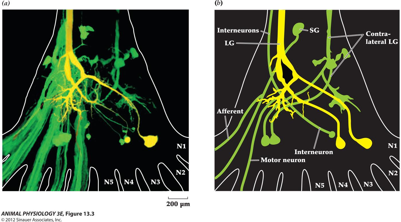
- The lateral giant neuron (LG) is dye-coupled with the green fluorescent neuron through Gap junction. LG was injected with large red and small green molecules.
2. Chemical Synapses
Chemical synapses can modify and amplify signals
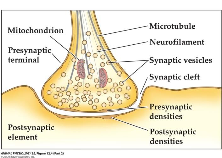
Chemical synapses have discontinuity due to the 20-30 nm synaptic cleft
Neurotransmitters are stored in synaptic vesicles with several thousand molecules per vesicle.
Both the pre and post synaptic membranes are thicker than elsewhere due to local protein aggregation.
**Active zone **– cytoplasmic side of the presynaptic membrane where synaptic vesicles release their neurotransmitter.
Postsynaptic density – thick membrane at the postsynaptic membrane containing neurotransmitter receptors and scaffolding proteins which organize these receptors and other proteins
The structure of a chemical synapse
The majority of excitatory synapses in a mammalian brain occur on dendritic spines
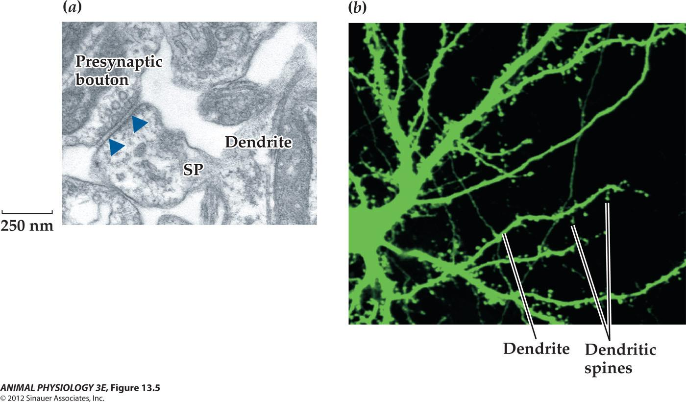
- Dendritic spines – small mushroom-shaped Protrusions and their shape and form remain malleable even in mature CNS
The function of a chemical synapse
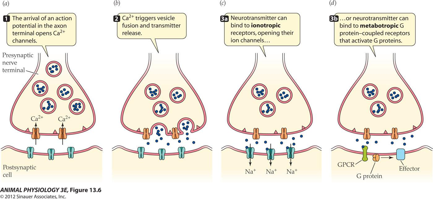
Neurotransmitter is released by calcium-dependent exocytosis – fusion of the Synaptic vesicles to the presynaptic membrane
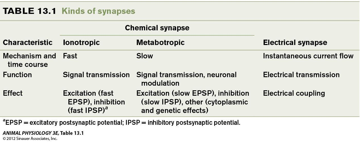
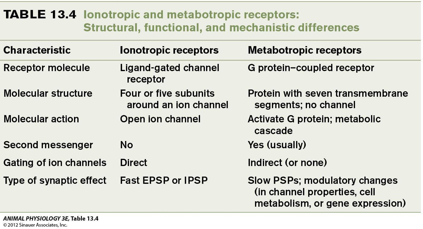
Neurotransmittor receptors:
- Ionotropic receptors (离子型受体)
- fast, permeability to ion changes Metabotropic
- receptors (代谢性受体)
- slow, long-lasting, triggering a signaling cascade
Advantage of chemical synapses
Chemical synapse is slower than the electrical synapse because the steps needed to release transmitters and receptor activation
- Fast ionotropic chemical synapses have a synaptic delay of 0.3 – 3 milliseconds, depending on species and temperature.
Advantages of chemical synapses:
- amplification of current flow–depending on the number of active zones and postsynaptic terminals.
- Both excitatory and inhibitory, while electrical synapse is always excitatory.
- Chemical synapse is one-way(单向的), while electrical synapses are two-way(双向的).
- Plasticity: Modifiable than the electrical synapses, use and circumstances make them stronger and disuse make them weaker. This plasticity is important for nervous system development and learning.
Synaptic potentials control neuronal excitability
Synaptic potential – a transitory graded change in the resting membrane potential in the postsynaptic cell caused by the burst of neurotransmitter diffuse across a synapse.
- Synaptic potential depolarizes the cell membrane is excitatory; one that hyperpolarizes the cell membrane is inhibitory.
Excitation: increase in generation of an action potential and increase the frequency of impulses.
Inhibition: decrease in the probability to generation of impulse of action potential and the frequencies of the impulses.
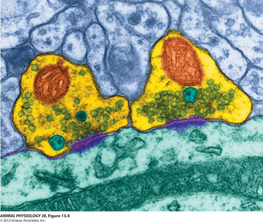
Excitatory and inhibitory postsynaptic potentials:
- synapses onto a spinal motor neuron exemplify functions of fast synaptic potentials
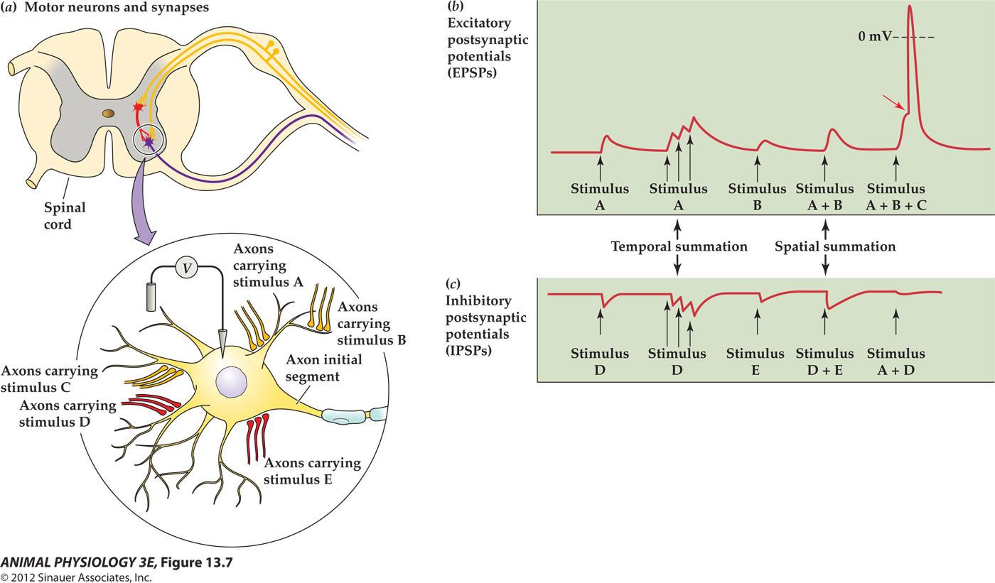
- Clusters of synapses: yellow color indicates excitatory Presynaptic terminals, red color indicates inhibitory Presynaptic terminals.
- Excitotaory postsynaptic potentials (EPSPs) Motor neuron membrane produces synaptic potentials that are graded depolarizations with a brief rising phase and an exponential decay over a time course of 10 – 20 ms.
- Simulation of excitatory presynaptic neurons elicits excitatory postsynaptic potentials (EPSPs), graded depolarization’s that depolarize Vm (membrane potential toward threshold
- IPSPs are hyperpolarizing, moving Vm away from threshold.
1. Neuronal Integration
Temporal summation(时间总和) (peripheral nerve A is stimulated repeatedly and rapidly and the resultant EPSPs combine in a process called temporal summation)
Spatial summation(空间总和) (stimulus A + B combine)
With sufficient presynaptic stimulation, EPSPs can summate temporally and spatially to the voltage threshold and cause the motor neuron to generate one or more action potentials.
A cat motor neuron receives input from 10,000 synaptic terminals and the moment-to-moment balance of EPSPs and IPSPs determines whether the motor neuron generates impulses or remain quiescent
A neuron’s output is not the same as its input, but is instead an integral function (整函数) of that input, a property called neuronal integration.
The major process underlying neuronal integration is
- The spatial and temporal summation of EPSPs and IPSPs.
- Spatial relationships of excitatory and inhibitory synapses to one another
- Their relative proximity to the site of impulse initiation – the initial segment of the axon
2. Summary
Summation of synaptic input is weighted by the electronic distance of the synapses from the axon hillock
Postsynaptic potentials are graded potentials
- Their spread is governed by the cable properties of the postsynaptic cell membrane.
- Axosomatic synapse(轴体突触) – a synapse located on the soma or cell body – very close to the axon initial segment
- Axodendritic synapse(轴树突触) – a synapse onto a dendrite
- The electrotonic length of motor neuron dendrites is estimated 1-2 lambda (so a synaptic potential at the tip of dendrites is decreased to 14 – 37% of the initial amplitude.
- Synapse closer to the axon hillock usually have more effect on the output of the postsynaptic cell that do synapses on the distal ends of dendrites
Neuromuscular synapse
1. Basic Structure and Mechanism
A vertebrate neuromuscular junction (Part 1) : an example of a fast chemical synaptic actions
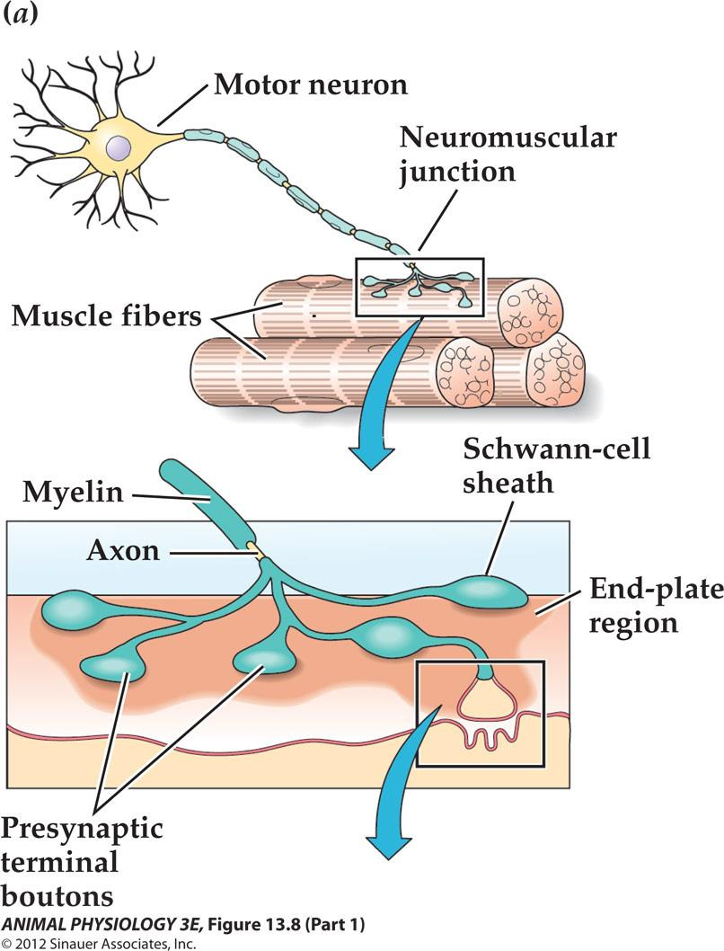
Each muscle fiber is innervated by Only one motor neuron.
Vertebrate neuromuscular junction – a model for fast chemical synaptic actions:
- Also called motor end-plate
- Anatomically simple, large synaptic response, microscopically visible,
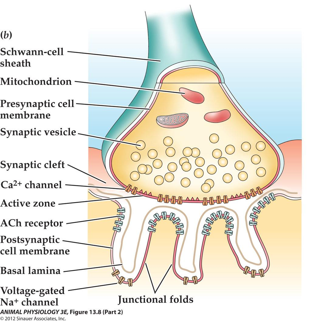
- Ligand-gated Ach receptors at the top part of the junctional fold
- Voltage-gated channels in the deep part of the fold
Summary of events in chemical synaptic transmission at the vertebrate neuromuscular junction
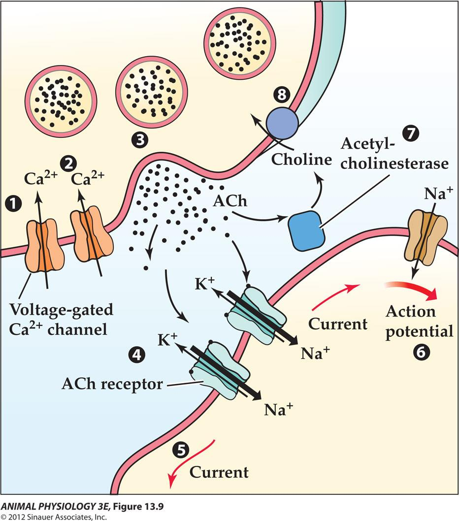
- An action potential depolarizes the axon terminal of the motor neuron.
- The depolarization opens voltage-gated Ca2+ channels.
- Depolarization of the terminal triggers vesicle exocytosis at an active zone, releasing acetylcholine(Ach).
- Ach diffuses rapidly across the synaptic cleft and binds to acetylcholine receptors at the postsynaptic membrane
- The receptor channel opens to allow Na”and K” ion flow, producing a depolarizing excitatory postsynaptic potential (EPSP). The EPSP spreads to depolarize nearby regions to threshold and triggers a muscle fiber action potential.
- The action potential propagates to all parts of the muscle fiber, eliciting contraction.
- Acetylcholinesterase hydrolyzes the acetylcholine into acetate and choline.
- Choline is actively transported back into the motor axon terminal to be resynthesized into acetyl
2. The Role of Permeability of Postsynaptic Membrane
Postsynaptic potentials result from permeability changes that are neurotransmitter-dependent and voltage-independent
Molecules that control permeability changes at fast synapses are ligand-gated!
Permeability of the postsynaptic is the result of binding of neurotransmitter, not to depolarization!
- An EPSP results from a simultaneous increase in the postsynaptic membrane’s permeability to Na and K , but the driving force for Na is higher and the driving force for K is smaller
- The ion flows through all the channels that open in response to the release of neurotransmitter is called synaptic current
Fast IPSPs can result from an increase in permeability to chloride
- Hyperpolarizing rather than depolarizing due to increased permeability to Cl-
- Mediated by two neurotransmitters: GABA (gamma-aminobutyric acid) or glycine
- GABA-containing neurons – vesicle fusion – GABA release – GABA fuse to the postsynaptic membrane – increase Clentry – hyperpolarize membrane and produce fast IPSPs
Presynaptic neurons release neurotransmitter molecules in quantal packets
- Cholinergic transmission (acetylcholine-mediated transmission) is a good general example
- Acetylcholine is made and stored in the presynaptic terminal
- Neurotransmitter release is quantal and vesicular
- Each quantum is equivalent to the contents of one synaptic vesicle of about 5000 molecules eachs
3. Comparison between Neuromuscular Synapse and CNS Synapse
A neuromuscular synapse, a CNS excitatory synapse, and a CNS inhibitory synapse
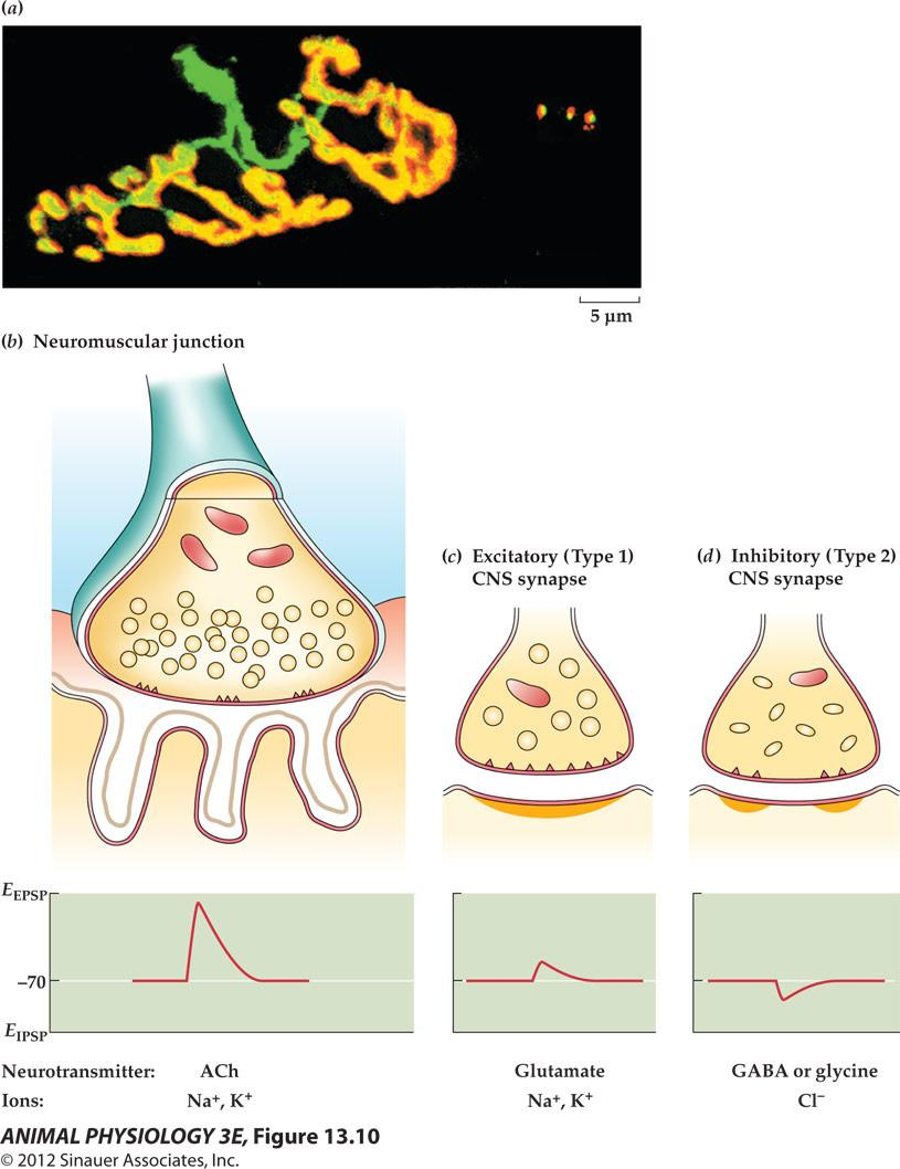
- CNS synapse
- Neuromuscular junction (green: presynaptic axon terminal; red: postsynaptic receptors; yellow: the synapses
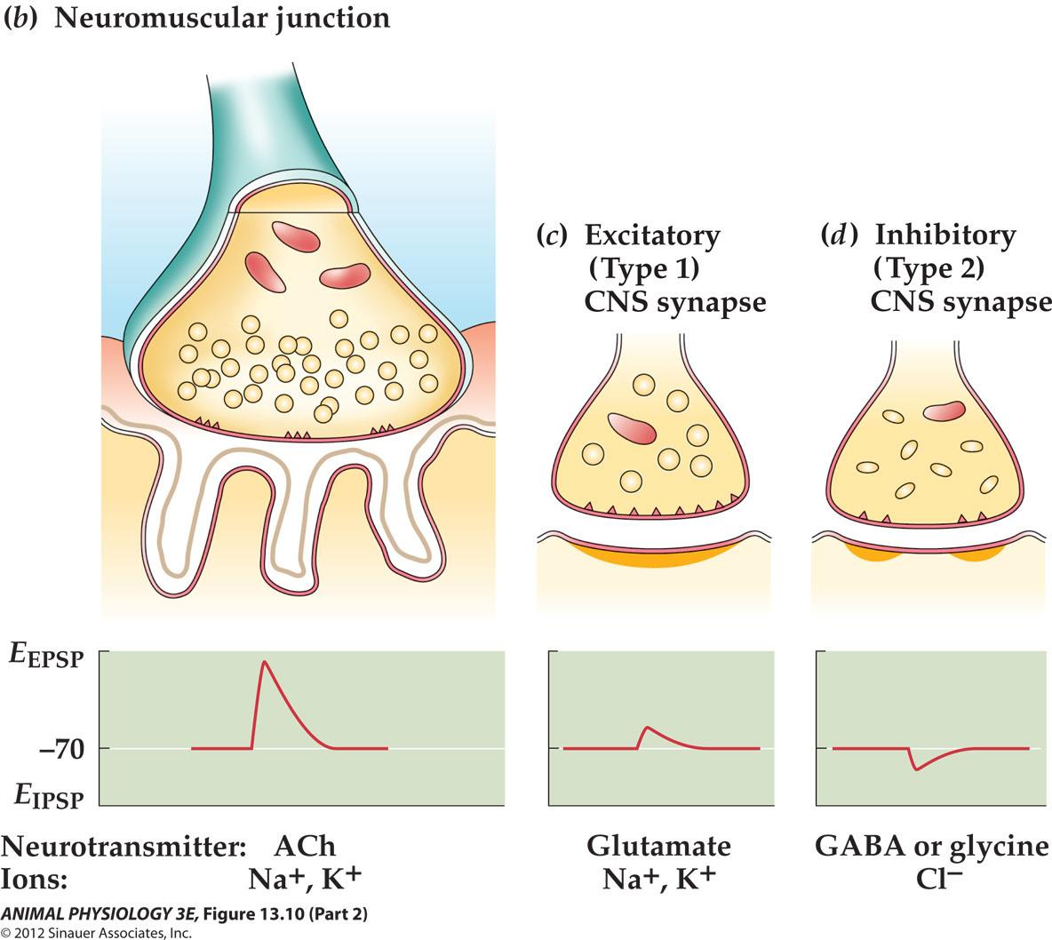
- Fast rise 1-2 ms And exponential return lasting 1020 ms
EPSPs between neurons resemble neuromuscular EPSPs but are smaller
- CNS synapses are integrating synapses (excitatory input from 20 to 50 neurons to be summate to depolarize the integrating neuron),
- while neuromuscular junction is a relay synapse (each presynaptic action potential produces a postsynaptic action potential)
4. Acetylcholine synthesis and breakdown
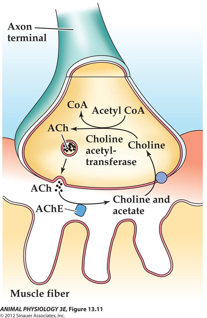
- Choline acetyltransferase is made in the cell body And the availability of choline is the limiting factor
- Choline is supplied by the blood and by choline uptake transporter at the presynaptic Membrane.
- Neurotransmitter release requires voltage-dependent calcium influx (bathing neuromuscular Junction in a calcium free saline will not elicit EPSP Because there is no exocytosis.
5. Spontaneous mEPSPs
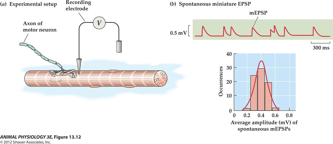
- A series of small depolarization with a shape similar to neuromuscular EPSP but are about 1/50th the amplitude (0.4 mV),
- Thus they are termed spontaneous miniature EPSPs (mEPSPs).
- A spontaneous, low-frequency release of Ach quanta-that is about 5000 molecules at a time Which is accounted for spontaneous mEPSPs.
Evoked miniature EPSPs
A neuromuscular EPSP evoked by a presynaptic impulse has an amplitude of 20 – 40 mV And requires the nearly simultaneous discharge of 100 to 300 quanta.
To determine if the release of Ach evoked by presynaptic depolarization is also quantal, researcher lowered Ca concentration in the bath and raised Mg which competitively inhibits the action of Ca
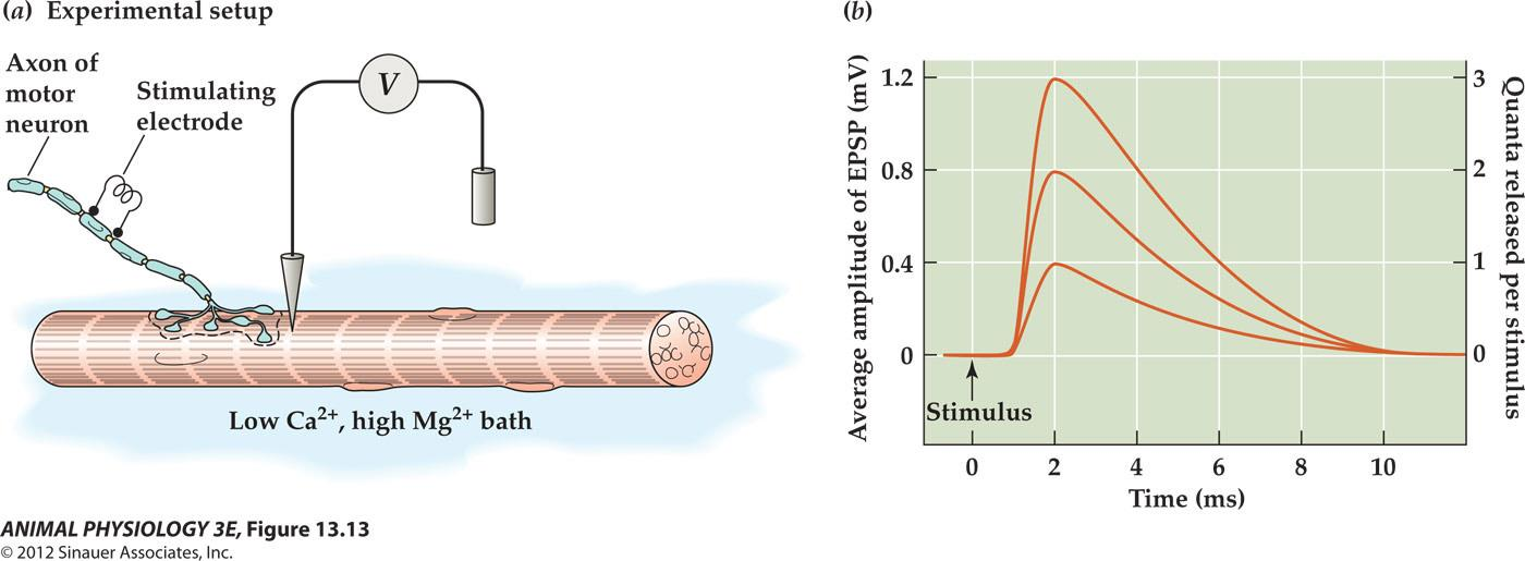
Evoked mEPSPs fall into amplitude classes that are multiples of the spontaneous mEPSPs.
6. Synaptic vesicles are cycled at nerve terminals in distinct steps
Vascular release hypothesis – vesicular exocytosis
Calcium ion triggers the fusion of the vesicular and presynaptic membranes
Two modes of fusion and subsequent retrieval of vesicular membranes: classical and kiss-and-run fusion
- Classical endocytosis: needs two proteins: clathrin and dynamin
- Kiss-and-run fusion: the docked vesicle opens only a pore to release neurotransmitter without completely becoming integrated into the terminal membrane
Synaptic vesicle recycling at the neuromuscular junction
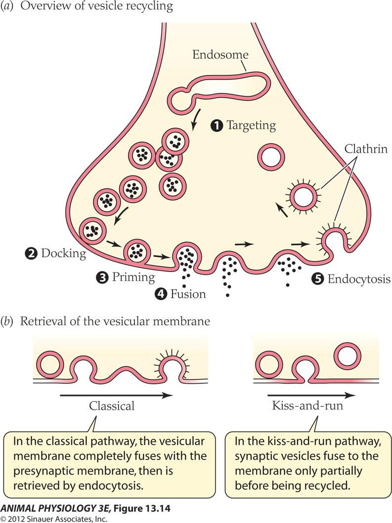
Stages of vesicular recycling:
- mobilization or targeting: at rest, synapsin attaches vesicles to the actin cytoskeleton
- phosphorylation of synapsin releases vesicles during mobilization to allow vesicles to move to the active zone
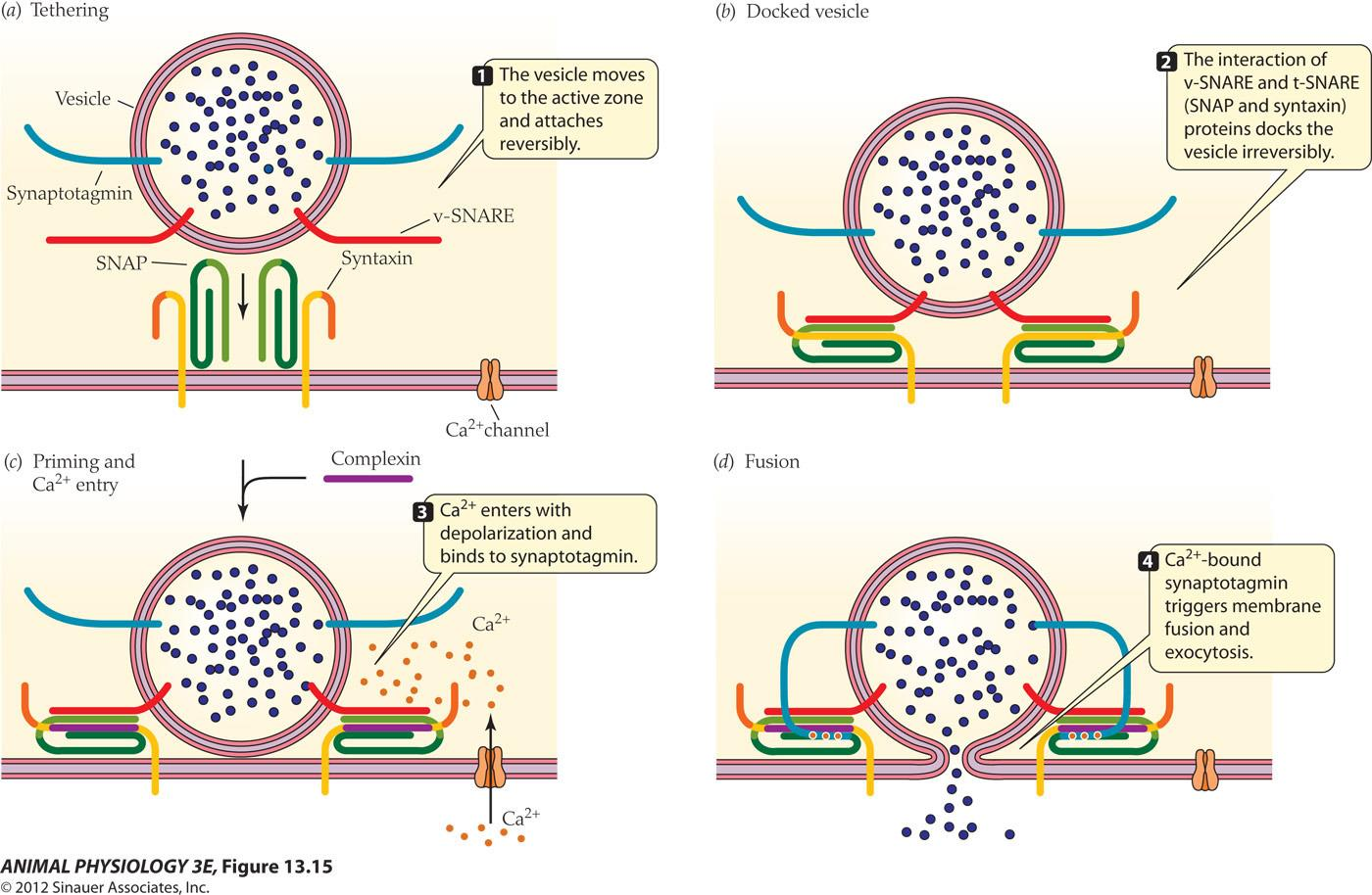
- vSNAREs intertwine with terminal-membrane t-SNAERs to hold the docked vesicle at the active zone
- Classical and kiss-and-run
Vesicular docking and fusion release neurotransmitters
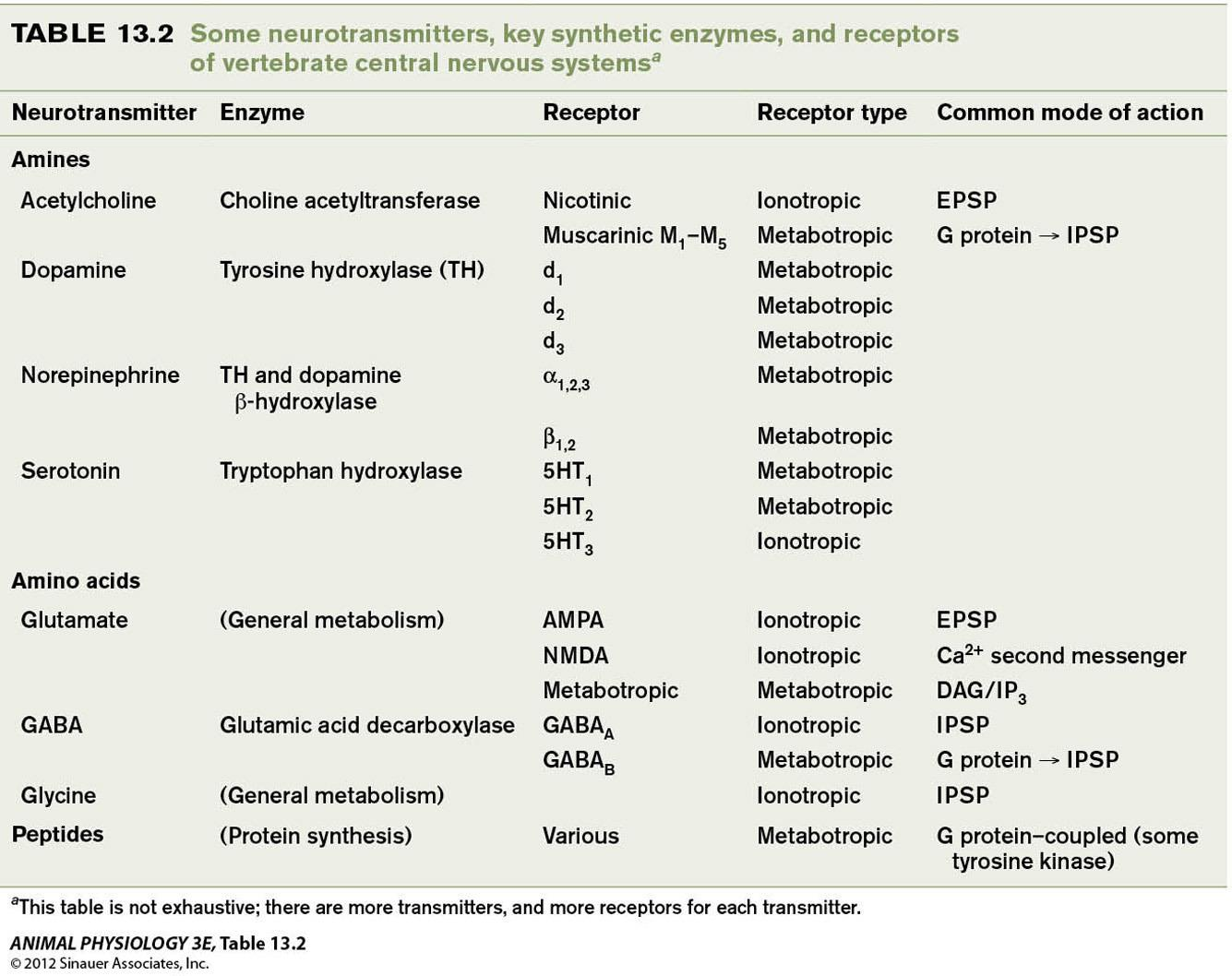
Neurotransmitters
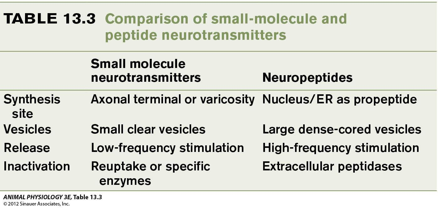
1. Neurotransmitters are of two general kinds
- Small molecule neurotransmitters (mostly amines and amino acids)
- Neuropeptides
- Cholinergic synapses are the best known because of the neuromuscular junction studies (acetylcholine as the neurotransmitter). 乙酰胆碱
- Adrenergic synapses are the next best known (noradrenaline as neurotransmitter).去甲肾上腺素
- A neuron may release more than one transmitters cotransmitters
2. Criteria required to be identified as a neurotransmitter
The candidate neurotransmitter must:
- be present in the presynaptic terminal along with its synthetic machinery
- Released upon stimulation
- Mimic the effect of presynaptic stimulation when added in extracellular fluid
- Mechanism of removal should exist
- Effects of drugs on transmission: curare is Ach receptor antagonist!
3. Termination of neurotransmitter action: enzymes and reuptake
Neurotransmitters are only active for a very short time (5 ms)
Acetylcholinesterase (AChE) located in the synaptic cleft acts very fast to digest Ach. (enzymatic destruction)
$$
\text{Ach} \xrightarrow{\text{AchE}} \text{Choline} + \text{acetate}
$$
(choline is returned back to the presynaptic terminal by a specific transporter)
The synaptic action of norepnephrine is terminated by reuptake of the neurotransmitter, rather than the enzymatic destruction.
Peptide neurotransmitters and small molecular neurotransmitter differ in synthesis, release and termination
Postsynaptic Receptors
1. Ach Receptors
Postsynaptic receptors for fast ionotropic actions: ligand-gated channels
- Ach receptors (nicotinic) are the best example of ionotropic receptors
The molecular structure and function of a ligand-gated channel, the nicotinic acetylcholine receptor:
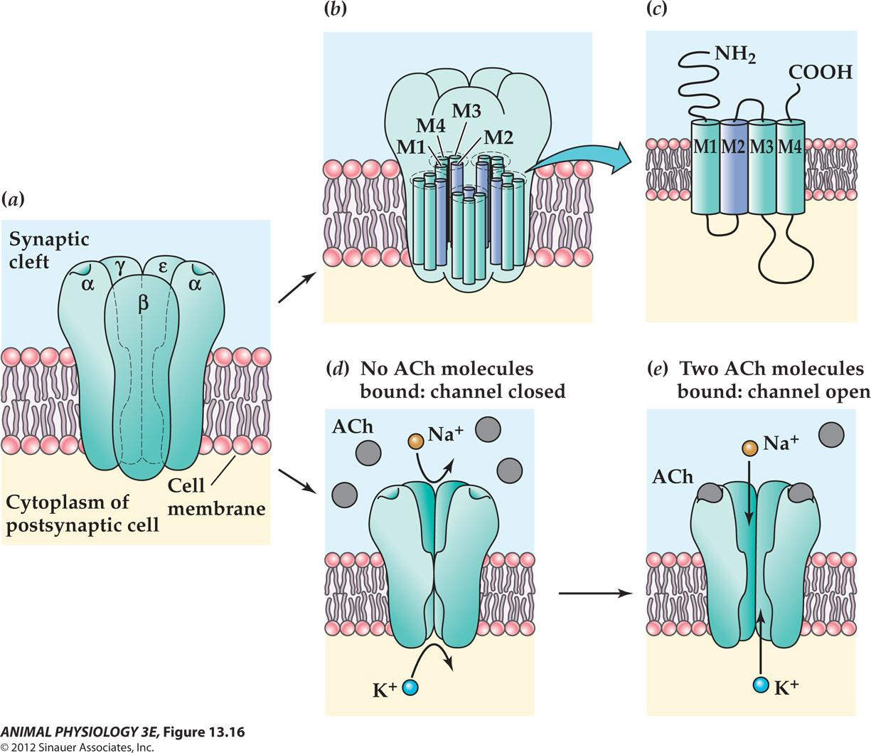
- Diameter of 0.6 nm
Patch-clamp recordings of acetylcholine receptor–channel currents
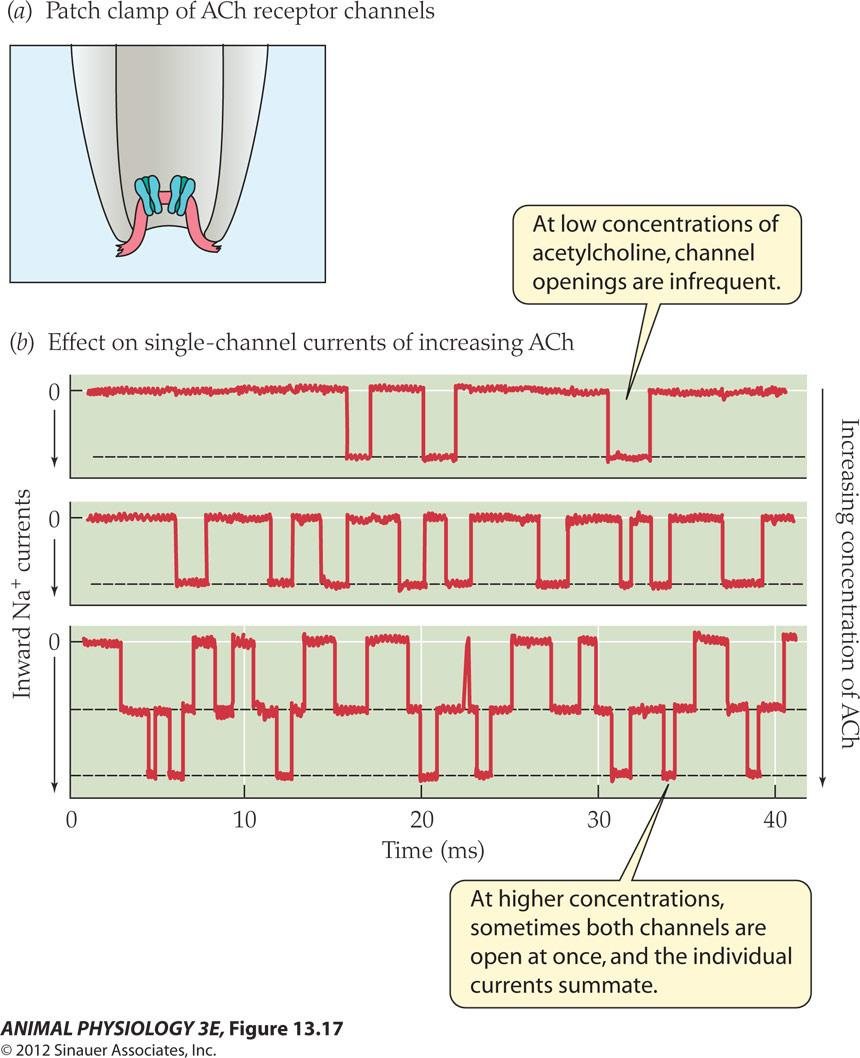
- At low concentrations of acetylcholine, channel openings are infrequent
- At higher concentrations, sometimes both channels are open at once, and the individual currents summate
The opening of the ligand-gated channel is an all-or-none phenomenon
Channel opening depends on the concentration of neurotransmitter at the receptor
The net ionic current through the Open channel provides that channel’s Contribution to a synaptic potential
The current through all open channels can be summated and constitute the synaptic currents
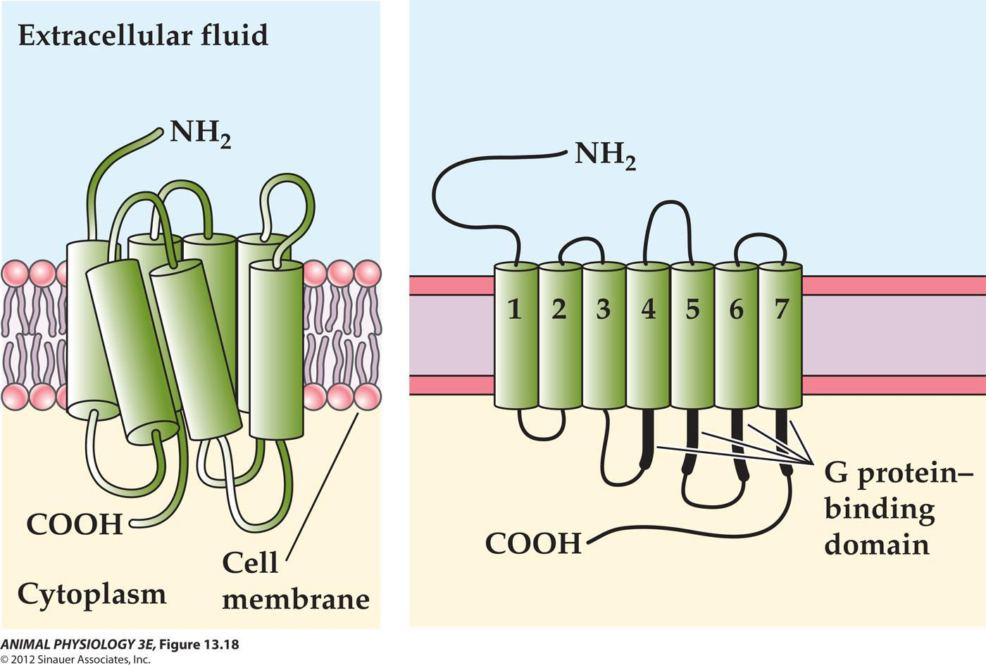
2. Postsynaptic receptors for slow metabotropic actions: G protein coupled receptors
Metabotropic receptors act via second messengers
GPCR has seven transmembrane segments
G protein–coupled receptors share a common structure:
3. Metabotropic receptors: cAMP as a second messenger
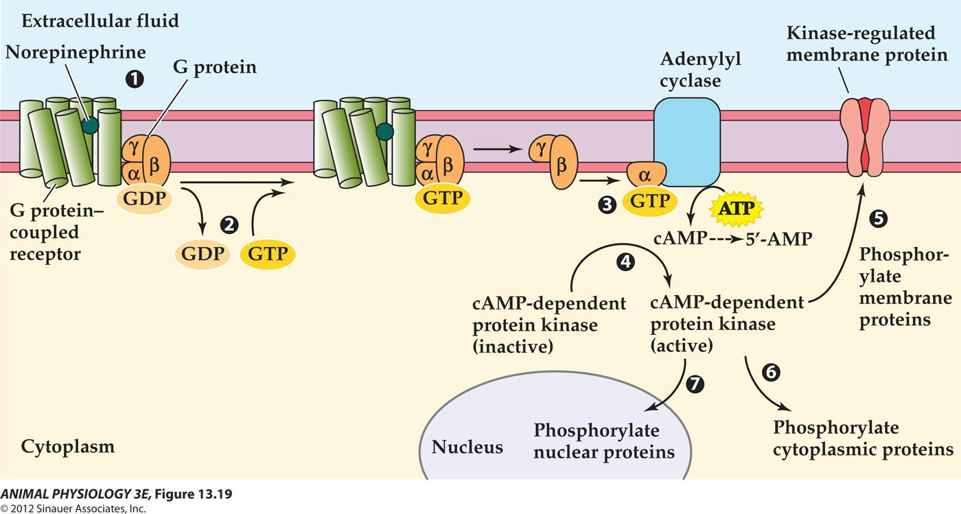
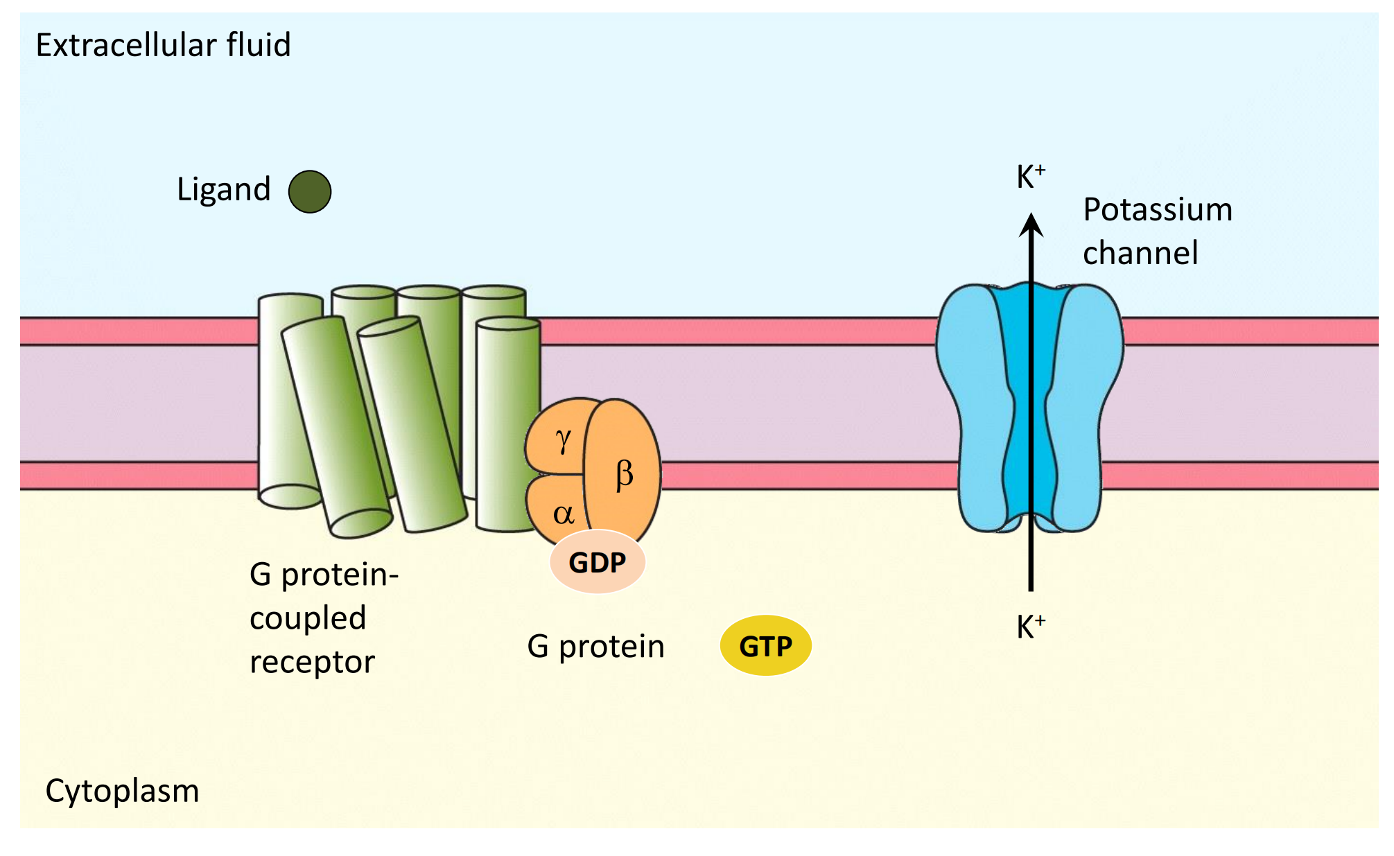
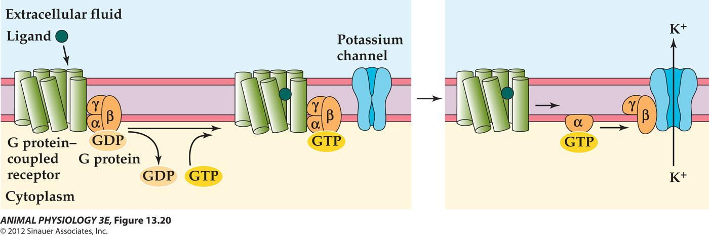
G proteins can themselves activate ion channels, without a second messenger
4. Metabolic receptors: Diacylglycerol and inositol trisphosphate are other second messengers
- Phospholipase C catalyzes the hydrolysis of PIP2 into IP3 and DAG
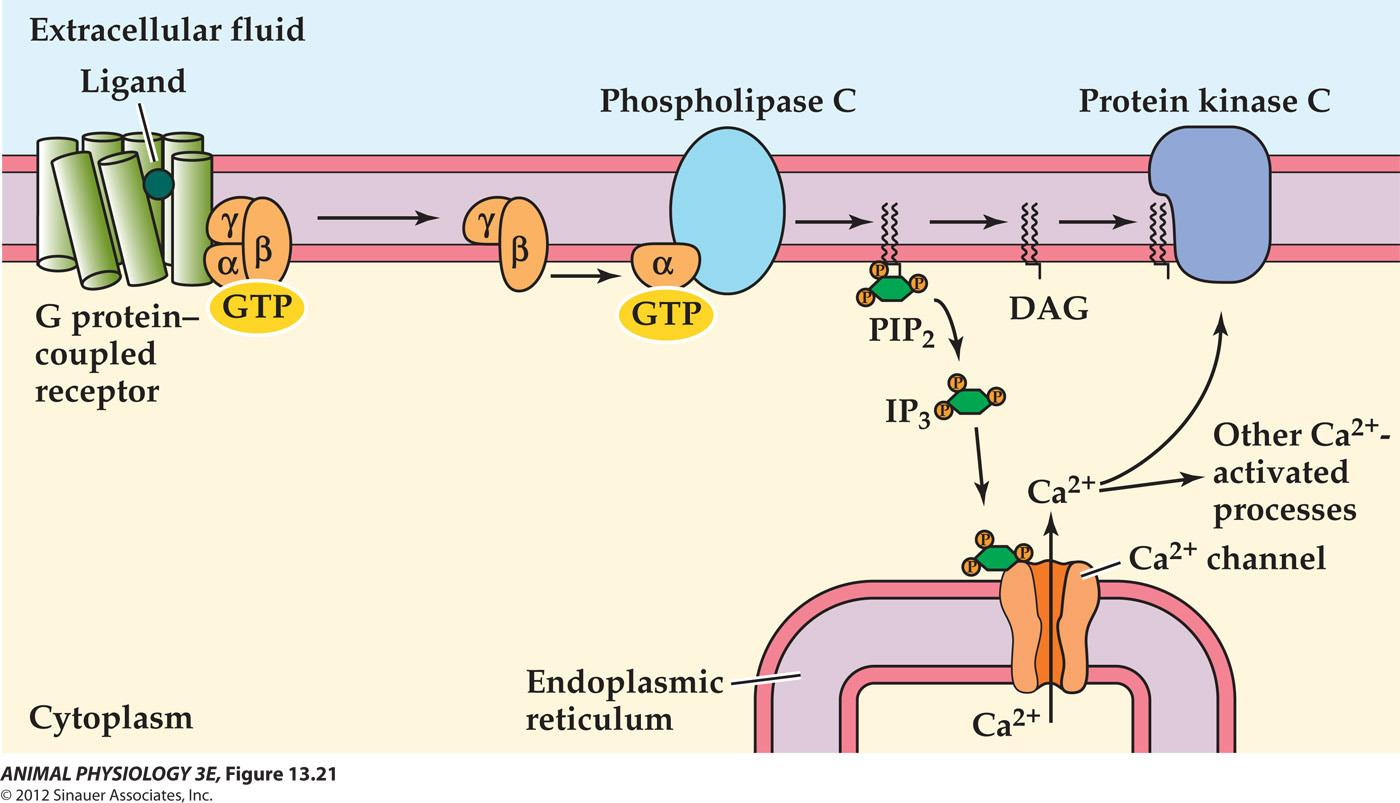
Together these metabotropic synaptic transmission can mediate relatively slow and long lasting effects
The second messenger action may underlie synaptic changes involved in learning and memory!
5. Metabotropic receptors plays a role in slow synaptic potentials
Permeability-decrease synaptic potentials – a slow EPSP results in ion permeability decrease due to the closing of a type of K+ channel
Presynaptic inhibition (PSI): specific inhibitory interaction in which one axon terminal ends on another axon (axo-axonal synapse). Presynaptic inhibition is more specific than the more common postsynaptic inhibition.
Synaptic plasticity: Synapse changes properties with time and activity
Neurotransmitter metabolism is regulated homeostatically (Ach level is stable during and after stimulation) – inhibitors to neurotranmitter synthesis and reuptake are used for psychiatric treatment (for example, selective serotonin reuptake inhibitors, resulting in increased availability of serotonin to postsynaptic receptors has been proven to be strikingly effective in treatment for depression and other mood disorders).
Learning and memory MAYBE based on synaptic plasticity
- (synaptic strength – changes that that have a suitable long time course of min, days and weeks. It is measured as the amplitude of a postsynaptic potential in response to a presynaptic potential. )
Synaptic facilitation and antifacilitation
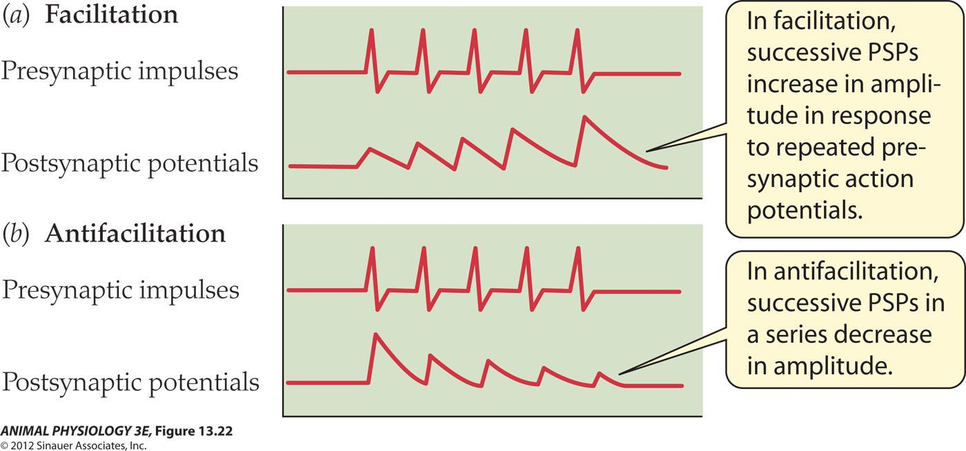
Both synaptic facilitation and antifacilitation result from changes in the amount of neurotransmitter liberated per presynaptic impulse, which is calcium dependent.
Tetanic stimulation of presynaptic neurons: stimulation by train of stimuli at a rate of 10-100 per second for several seconds
LTP and LDP
Posttetanic potentiation: Extended enhancement of synaptic response .
Long term potentiation: long-lasting enhancement of synaptic transmission following intense stimulation [ or long term depression (LDP)].
LTP has been reported in the hippocampus and cerebral cortex of the vertebrate brain, regions important in learning and memory.
1. Habituation and sensitization in Aplysia gill withdrawal
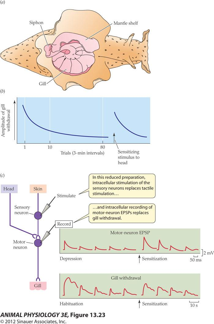
Eric Kandel – Nobel-Prize winning neurobiologist Mapped the neural circuit of the gill-withdrawal reflex And determined the synaptic locus of the habituation and sensitization
Habituation: the decrease in intensity of a reflex response to a stimulus when the stimulus is presented repeatedly
Sensitization: the prolonged enhancement of a reflex response to a stimulus resulting from the presentation of a second stimulus.
Both are simple forms of learning – modification of behavior with experience
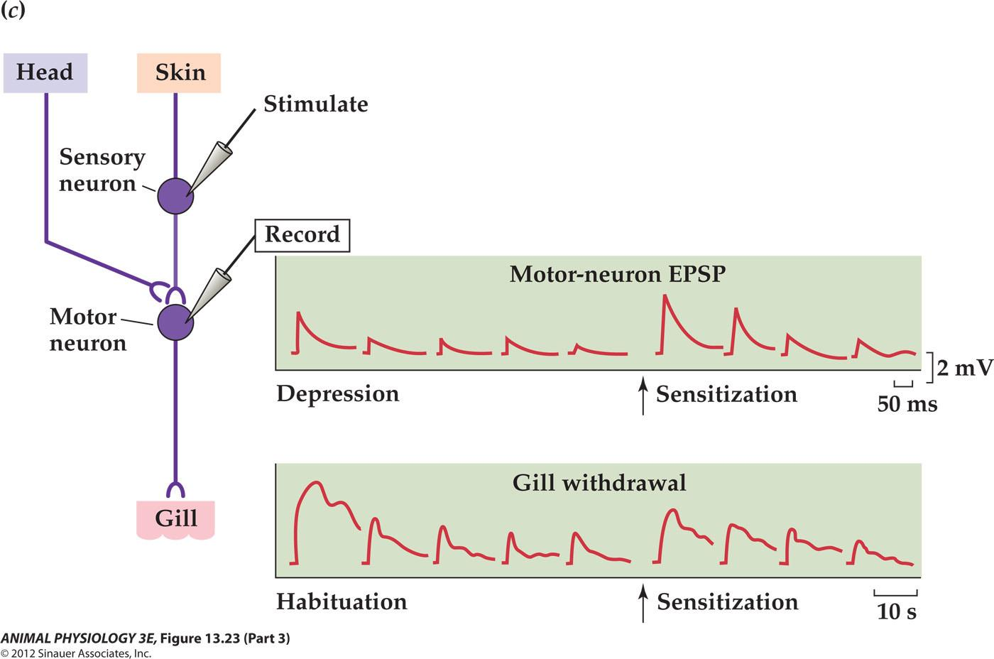
- How is neurotransmitter release diminished during habituation and increased by sensitization?
- Progressive and long lasting inactivation of calcium channels during habituation
- And increased calcium influx during presynaptic Facilitation at the axo-axonal synapse underlies sensitizations
A model of Aplysia sensitization
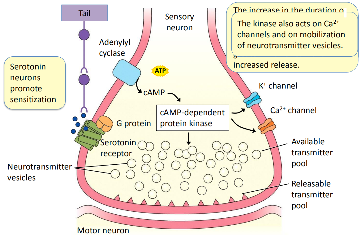
- Serotonin neurons promote sensitization
- The increase in the duration of the action potential increases the time during which Ca2+ channels can open, leading to a greater influx of Ca2+ and increased release
- The kinase also acts on Ca2+ channels and on mobilization of neurotransmitter vesicles
Both synaptic facilitation and antifacilitation result from changes in the amount of neurotransmitter liberated per presynaptic impulse. These changes are calcium dependent and the complete mechanism is incompletely known.
Facilitation is often pronounced after tetanic stimulation (强直刺激) of presynaptic neurons (a rain of stimulation at a rate of about 10 – 100 per second for several seconds). The extended enhancement of synaptic response is termed posttetanic potentiation.
Posttetanic potentiation means synaptic efficacy can change with use and these changes can be long-lasting
Long-term potentiation occurs in hippocampus and cerebral cortex, regions implicated in learning and memory
2. Long-term potentiation (LTP) in the hippocampus
LTP is a long lasting enhancement of synaptic transmission following intense stimulation
LTP in the brain involves changes in synapse strength (neurons that fire together wire together – Hebbian synapse, or Hebbian Theory)
Hippocampal LTP is established depending on the NMDA receptor and AMPA receptor.
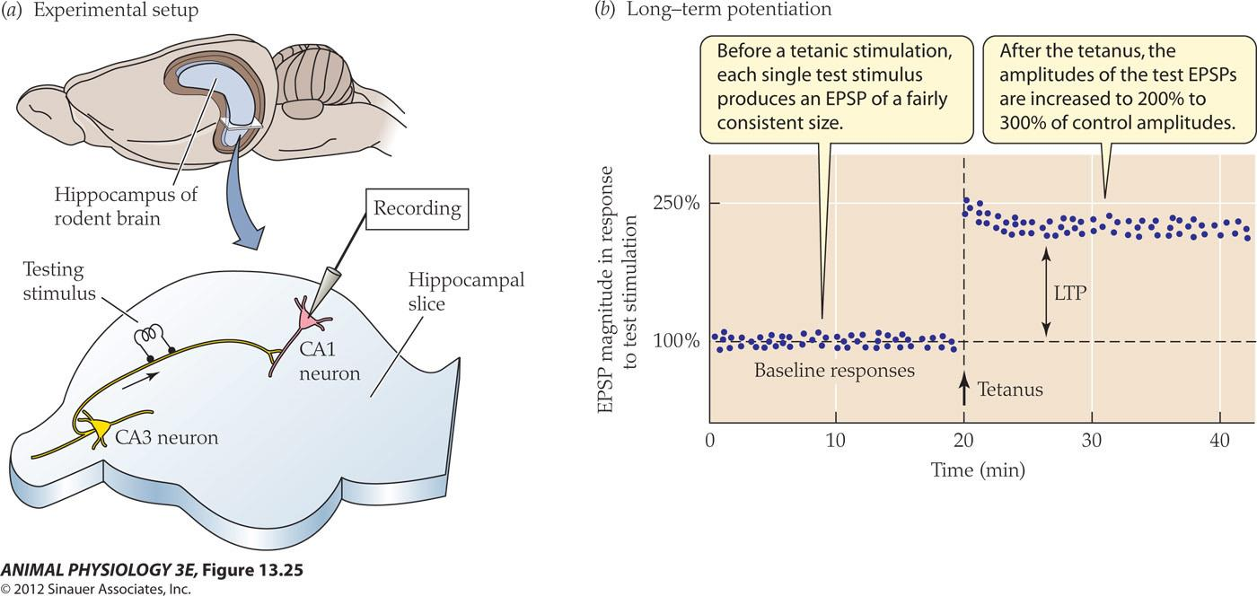
- Before a tetanic stimulation each single test stimulus produces an EPSP of a fairly Consistent size
- After the tetanus, the amplitudes of the test EPSP are increased to 200% to 300% of control amplitudes
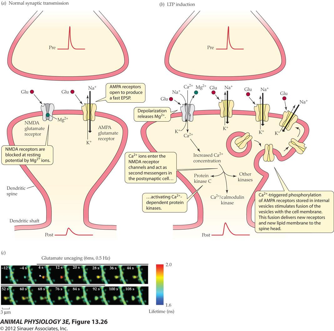
Induction and maintenance of LTP in the hippocampus:
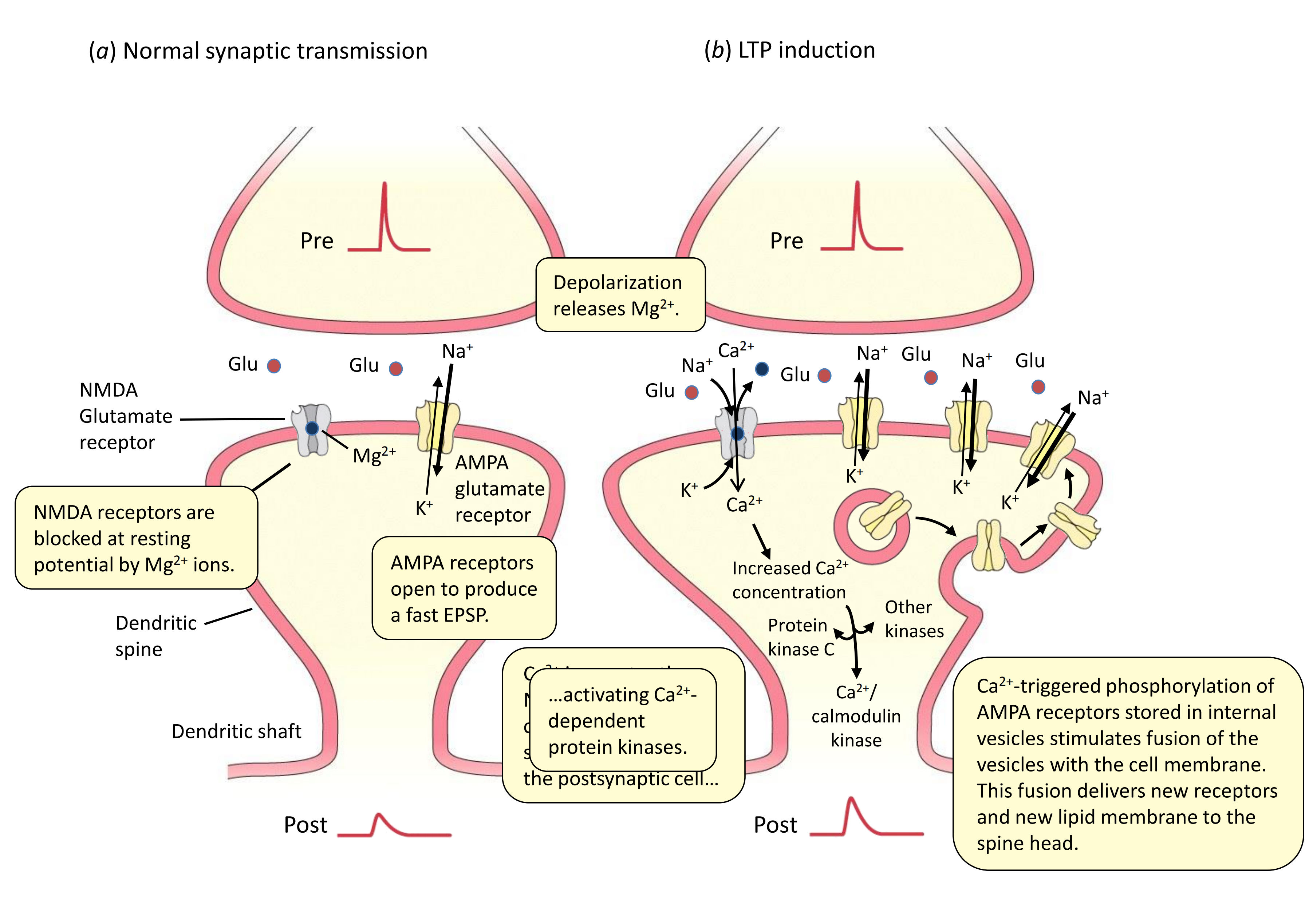
- Normal synaptic transmission
- Depolarization releases Mg2+
- NMDA receptors are blocked at resting potential by Mg2+ ions
- AMPA receptors open to produce a fast EPSP.
- LTP induction
- Depolarization releases Mg2+
- Ca2+ ions enter the NMDA receptor channels and act as second messengers in the postsynaptic cell…
- NMDA receptor channels and act as second messenger
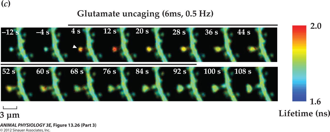
- Ca2+ -triggered phosphorylation of AMPA receptors stored in internal vesicles stimulates fusion of the vesicles with the cell membrane. This fusion delivers new receptors and new lipid membrane to the spine head.
Enduring changes in synaptic strength is caused by postsynaptic AMPA-type ionotropic glutamate receptors, which increase the amplitude of synaptic responses
 3. Long term depression (LDP) and LDP
Also mediated by NMDA receptors
Involving the removal of AMPA receptors from the postsynaptic membrane
4. Long term memories
Long term memories can involve changes to the physical structures of neurons
- LTP lasts for hours and weeks, some new genes and proteins are made, at the synapse and nucleus
- The excitatory synapses enlarge in response to local release of glutamate
- What is the character of these proteins and how are they transported to the correct synapses where the signals began? These are significant questions for the next generation of neuroscientists!
Repeated imaging of dendrites through cranial windows in living rodents
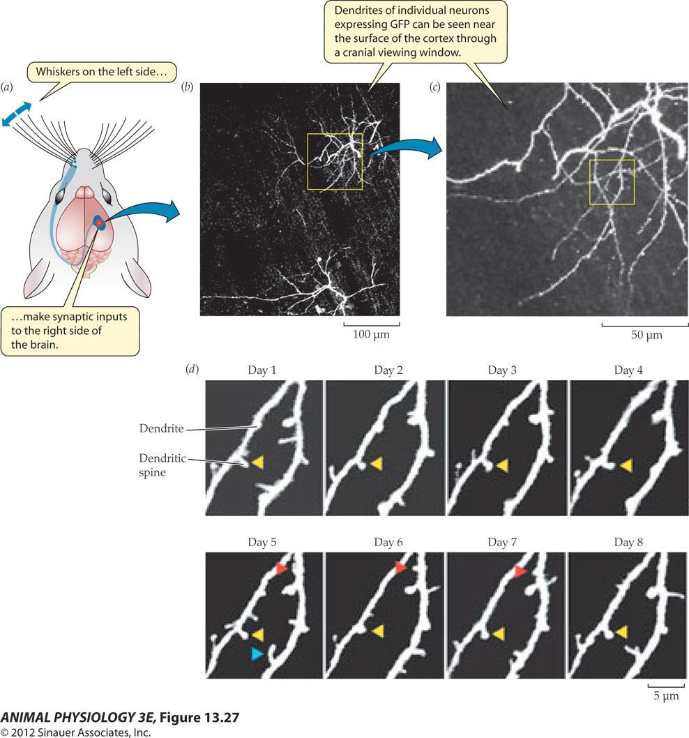
- Repeated imaging through a cranial window reveals Transient (blue), semistable (red) and stable (yellow) protrusions.
5. Long-term potentiation is a necessary component of learning
The existence of LTP DOES NOT prove that it is involved in learning and memory – only that it could be!
Knockout NMDA receptors, CaMK kinase and other kinases disrupts both LTP and spatial learning in a water maze. Same is true when AMPA receptor insertion into the postsynaptic membrane is disrupted
The most definitive answer comes from a relatively recent study of over-expressing NMDA receptors – Doogie mice – produced by Joe Tsien at Preston University. These mice performed several learning tasks better than normal mice.