L03 Bacterial Cell Structure
一、A Typical Bacterial Cell
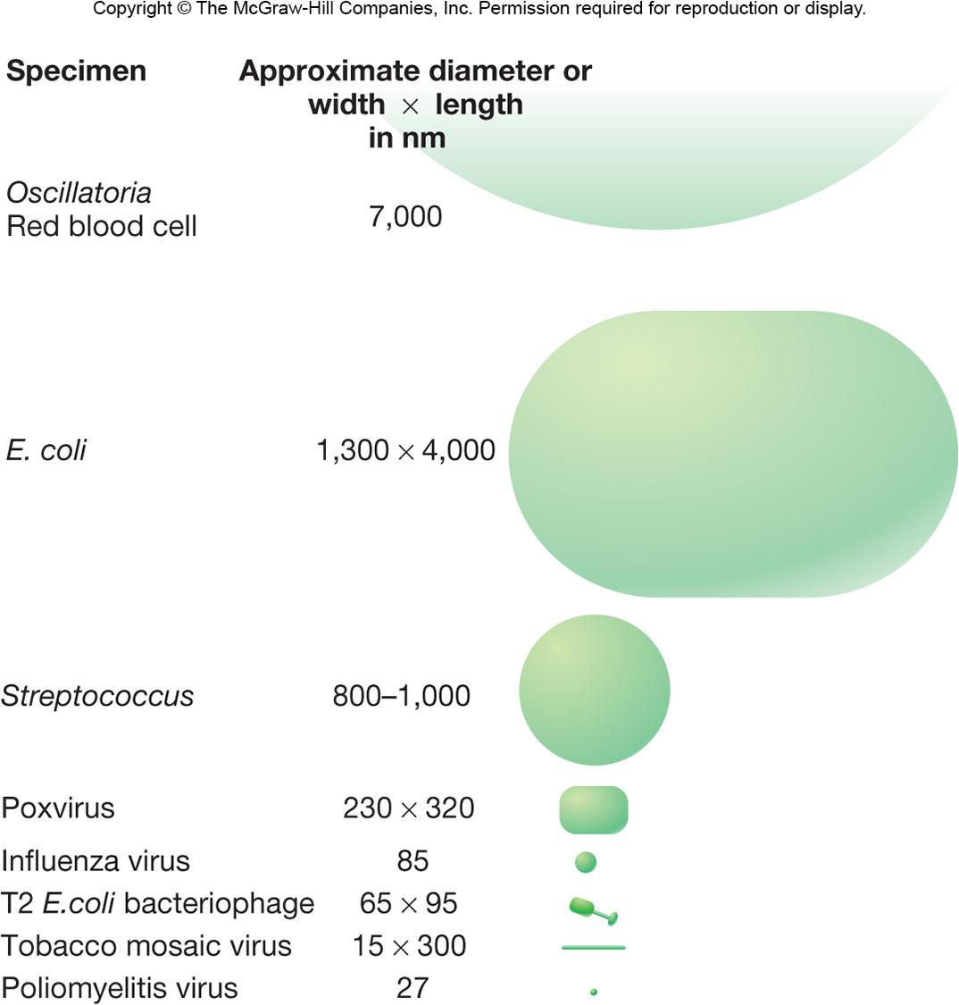
Size
The size of bacteria varies.
smallest : 0.3 μm (Mycoplasma)
average rod: 1.1-1.5 x2-6 pm (E.coli)
very large 一 600 x 80pm Epulopiscium fishelsoni
Shape and Arrangement
cocci and rod are most common, but have many other shapes.
怎么保持两个一组的?细胞膜表面的蛋白质?蛋白质位置?有什么用?提高物质交换的效率?
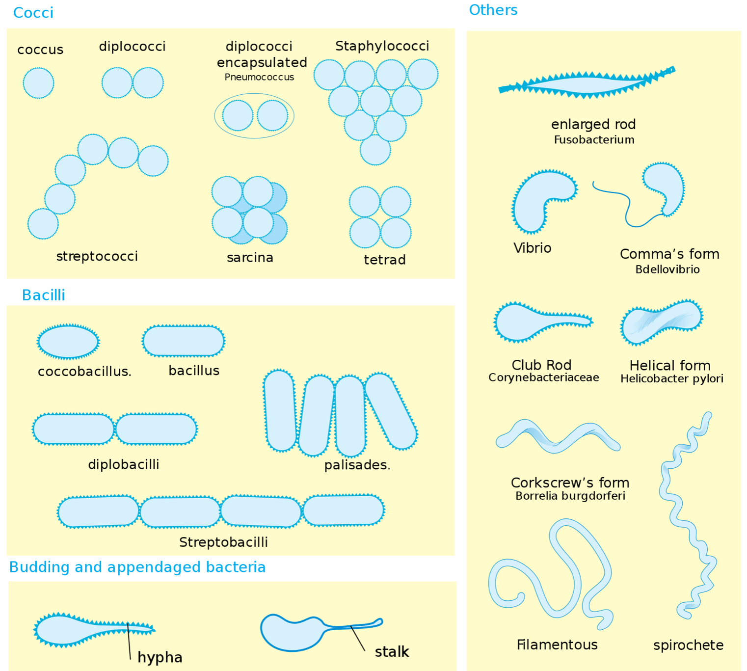
1. Cocci (s., coccus,球菌) – spheres
diplococci 双球菌
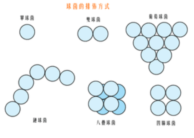
双球菌(拉丁语:diplococcus,复数diplococci)是球菌的一类,其细胞沿一平面分裂,而子细胞成双排列。代表种类有脑膜炎双球菌(Neisseria meningitidis)、淋球菌(Neisseria gonorrhoeae)等。
tetrads 四联球菌
四联球菌由四个球菌呈正方形排列
sarcinae 八叠球菌
与sarcina等同
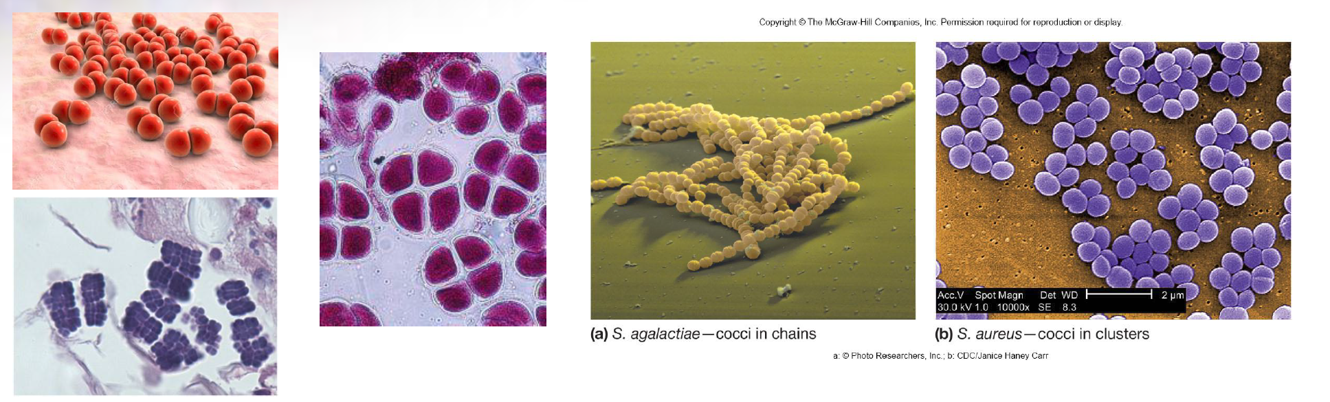
streptococci 链球菌
链球菌(Streptococcus)是一类球形的革兰氏阳性细菌,属于厚壁菌门的一个属。这些细菌细胞分裂时总是沿一个轴,所以通常成对或者链状的。因为这些特征,他们被称作“链球菌”,区别于可以沿多个轴分裂而形成一团细胞的“葡萄球菌”(Staphylococcus)。链球菌属包含了很多个种,其中多数是在人和动物表皮,呼吸道等处的共生菌(commensal flora), 也有对人类有益的菌种如嗜热链球菌(Streptococcus thermophilus),但其中也有相当数量的致病菌种.
staphylococci 葡萄球菌
葡萄球菌(学名:Staphylococcus)是一群革兰氏染色阳性球菌,因常常堆聚成葡萄串状而得名。在生物学分类上,葡萄球菌是芽孢杆菌目的一个属。多数葡萄球菌为非致病菌,少数可致病。有些菌种为人类皮肤和黏膜上的正常菌种,不会产生芽孢,夹膜不明显,无鞭毛和运动性。
代表种有金黄色葡萄球菌(S. aureus)、白色葡萄球菌(S. albus)、柠檬色葡萄球菌(S. citreus)等。
2. bacilli (s., bacillus芽孢杆菌属) – rods
芽孢杆菌属(Bacillus)属于革兰氏阳性菌,包括模式生物枯草芽孢杆菌和著名的致病菌炭疽杆菌等。此属细菌中有专性需氧菌或兼性厌氧菌,他们遍布于各种环境,有些独立存在亦有些会寄生于其他生物。在不适合生存的情况下,此属细菌可以转化为内生孢子(endospore)以进入休眠状态,此休眠状态可以持续很久。
在芽孢杆菌属中,其中枯草杆菌可以作为科学研究用的模式生物,还有可致病的炭疽杆菌和蜡样芽孢杆菌。
Coccobacillus 球杆菌
球杆菌(英语:coccobacillus 复数时:coccobacilli)是一种形状介于球菌、杆菌之间的微生物。它的形状是非常短的杆形,因而常常被误认为球菌。流感嗜血杆菌,阴道加德纳菌和沙眼衣原体均为球杆菌。Aggregatibacter actinomycetemcomitans菌是一种普遍存于牙龈菌斑上的革兰氏阴性菌,它也是球杆菌的一员。同属球杆菌的不动杆菌菌株可在固体培养基上生长。可诱发百日咳的百日咳杆菌同样是球杆菌。在医学上具有重要地位、能诱发布氏杆菌病的布氏杆菌,以及同样在医学上具有重要地位、在第三世界国家常见的能诱发软下疳的、属于革兰氏阴性菌的杜克雷嗜血杆菌也是球杆菌家族的一员。
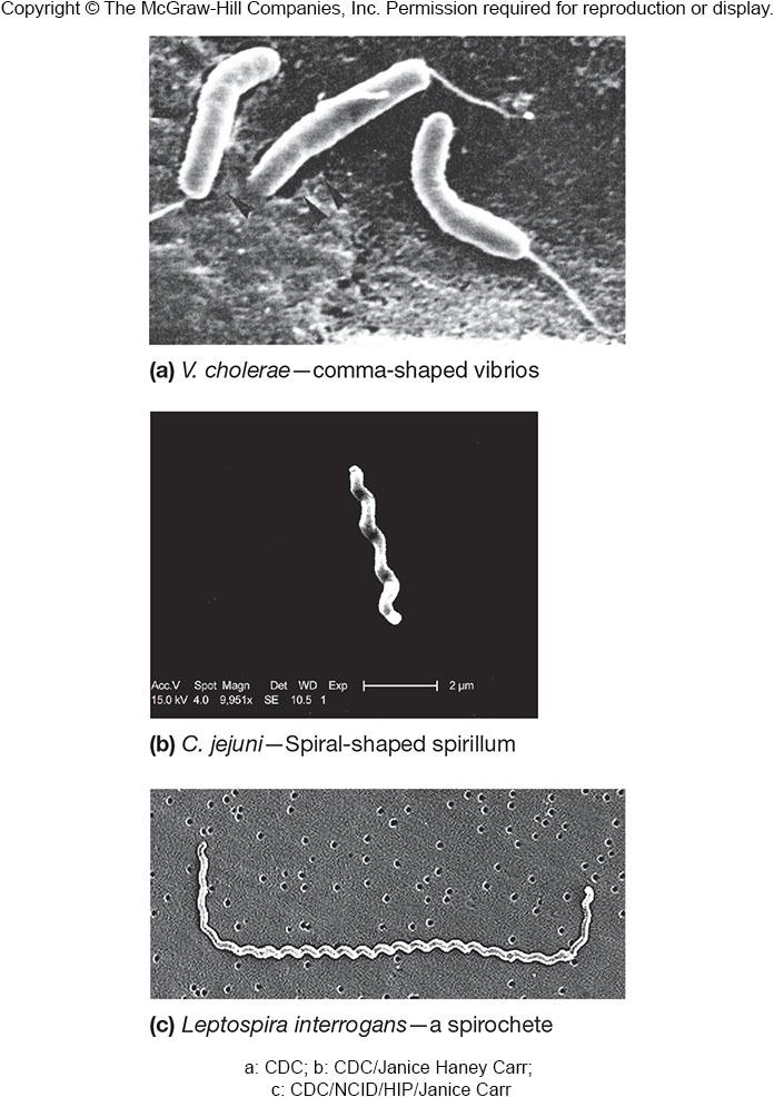
vibrios 弧菌
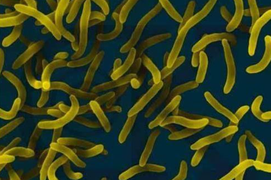
弧菌菌体只有一个弯曲,呈弧状或逗点状。如霍乱弧菌。
spirilla 螺旋菌
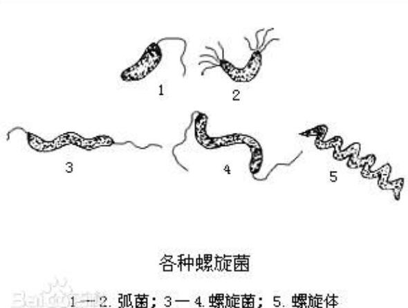
vibrios和spirilla的结构并不是flexible的,而螺旋体是flexible
spirochetes 螺旋体
螺旋体(spirochete)是一类细长、柔软、弯曲呈螺旋状、运动活泼的原核细胞型微生物。在生物学位置上介于细菌与原虫之间。螺旋体在自然界中分布广泛,常见于水、土壤及腐败的有机物上,亦有的存在人体口腔或动物体内。
3. Others
mycelium(菌丝体)
Some bacteria can be thought of as multicellular.
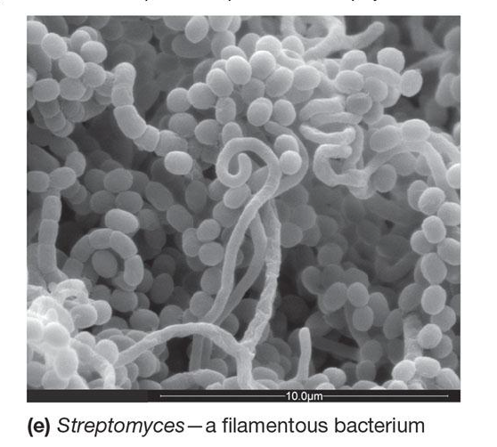
Actinomycetes(放线菌) typically form long filaments called hyphae(hypha的复数,菌丝). The hyphae may branch to produce a network called a mycelium, and in this sense, they are similar to eukaryotic filamentous fungi.
pleomorphic
这种细菌being variable in shape and lacking a single, characteristic form.
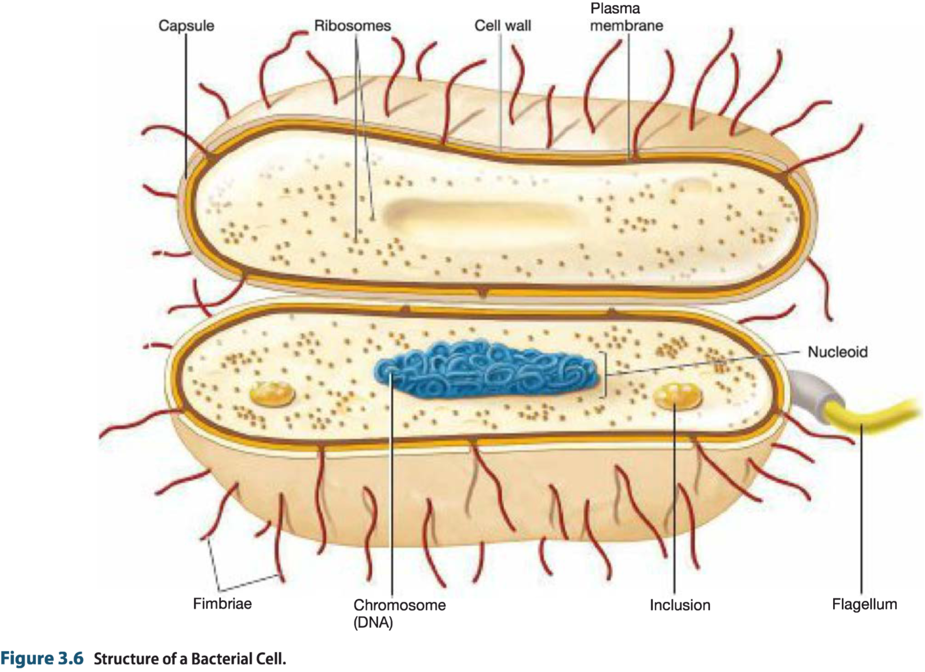
二、Bacterial Cell Organization(Bacterial Cell Envelope)
Cell envelope: The cell envelope is defined as the plasma membrane and all the surrounding layers external to it.
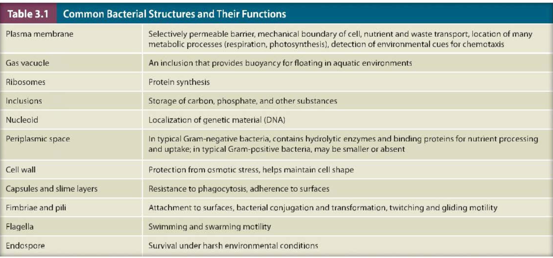
| Plasma membrane | Selectively permeable barrier, mechanical boundary of cell, nutrient and waste transport, location of many metabolic processes (respiration, photosynthesis), detection of environmental cues for chemotaxis |
|---|---|
| Gas vacuole | An inclusion that provides buoyancy for floating in aquatic environments |
| Ribosomes | Protein synthesis |
| Inclusions | Storage of carbon, phosphate, and other substances |
| Nucleoid | Localization of genetic material (DNA) |
| Periplasmic space | In typical Gram-negative bacteria, contains hydrolytic enzymes and binding proteins for nutrient processing and uptake; In typical Gram-positive bacteria, may be smaller or absent |
| Cell wall | Protection from osmotic stress, helps maintain cell shape |
| Capsules and slime layers | Resistance to phagocytosis, adherence to surfaces |
| Fimbriae and pili | Attachment to surfaces, bacterial conjugation and transformation, twitching and gliding motility |
| Flagella | Swimming and swarming motility |
| Endospore | Survival under harsh environmental conditions |
Note that no single bacterium possesses all of these structures at all times. Some are found only in certain cells in certain conditions or in certain phases of the life cycle.
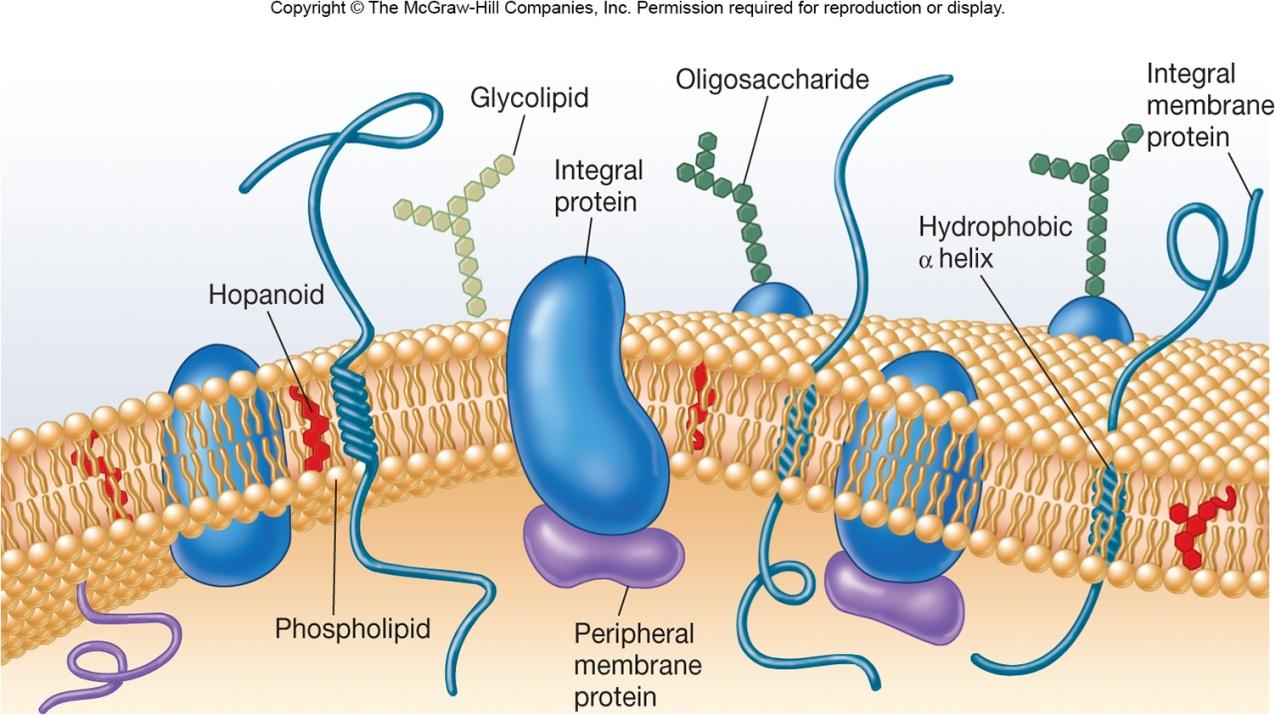
Bacterial Plasma Membranes
Encompasses the cytoplasm
Selectively permeable barrier
Interacts with external environment
- receptors for detection of and response to chemicals in surroundings
- transport systems
- metabolic processes
1. Membrane Proteins
- Integral membrane proteins(整合周蛋白,集成膜蛋白,内嵌膜蛋白):An integral membrane protein (IMP) is a type of membrane protein that is permanently attached to the biological membrane.
- Peripheral membrane proteins (外周膜蛋白):Peripheral Membrane Proteins are membrane proteins that adhere only temporarily to the biological membrane with which they are associated. These proteins attach to integral membrane proteins
- 跨膜蛋白(transmembrane protein,TP):跨膜蛋白是一种贯穿生物膜两端的蛋白。许多跨膜蛋白的功能是作为信道或“装载码头”来实施拒绝或允许某种特定的物质跨过生物膜的运输。所有的TP都是IMP
2. Fluid Mosaic Model of Membrane Structure
lipid bilayers with floating proteins
- amphipathic lipids
- polar ends (hydrophilic 一 interact with water)
- non-polar tails (hydrophobic - insoluble in water)
- Membrane proteins
3. Bacterial Lipid
Bacterial membranes lack sterols but do contain sterol-like molecules, hopanoids
- stabilize membrane
- found in petroleum
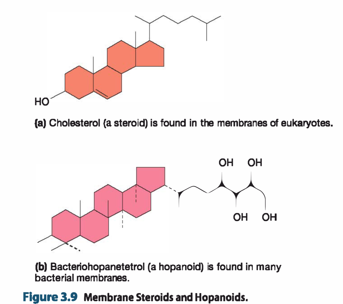
Bacterial membranes usually differ from eukaryotic mem branes in lacking sterols(甾醇类;固醇类) (steroid-containing lipids,含类固醇的脂质) such as cholesterol
However, many bacterial membranes contain sterol-like molecules called hopanoids(藿烷类化合物)
hopanoids have contributed significantly to the formation of petroleum(石油).
4. Uptake of Nutrients
Some nutrients enter by passive diffusion.
Microorganisms use transport mechanisms:
- Facilitated Diffusion—all microorganisms
- Active Transport—all microorganisms
- group translocation 一 Bacteria and Archaea
- endocytosis 一 Eukarya only
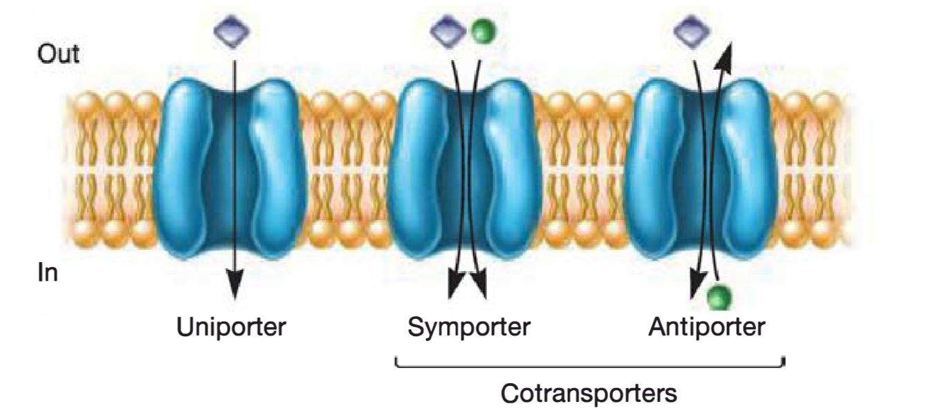
Primary active transport 原发性主动运输
直接利用ATP裂解所产生的能量进行物质运输 without modifying them
Primary active transporters are uniporters(单向运输载体); that is, they move a single molecule across the membrane
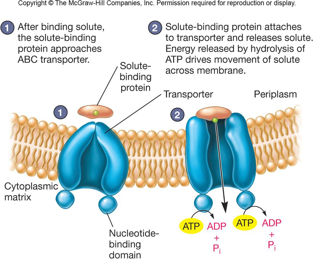
(1) ABC Transporters
ATP-binding cassette transporters (ABC transporters) are important primary active transporters. It is observed in Bacteria, Archaea, and eukaryotes
Primary active transporters use ATP
ABC transporters Consist of :
- 2 hydrophobic membrane spanning domains
- 2 cytoplasmic associated ATP-binding domains
- Substrate binding domains
Secondly active transport 继发性主动运输
继发性主动运输利用势能进行运输,在这个过程中也不会对所运输的物质进行修饰
Secondary active transporters include the major facilitator superfamily (MFS,转运蛋白超家族) proteins.
The lactose permease of E. coli is a well-studied secondary active transporter.
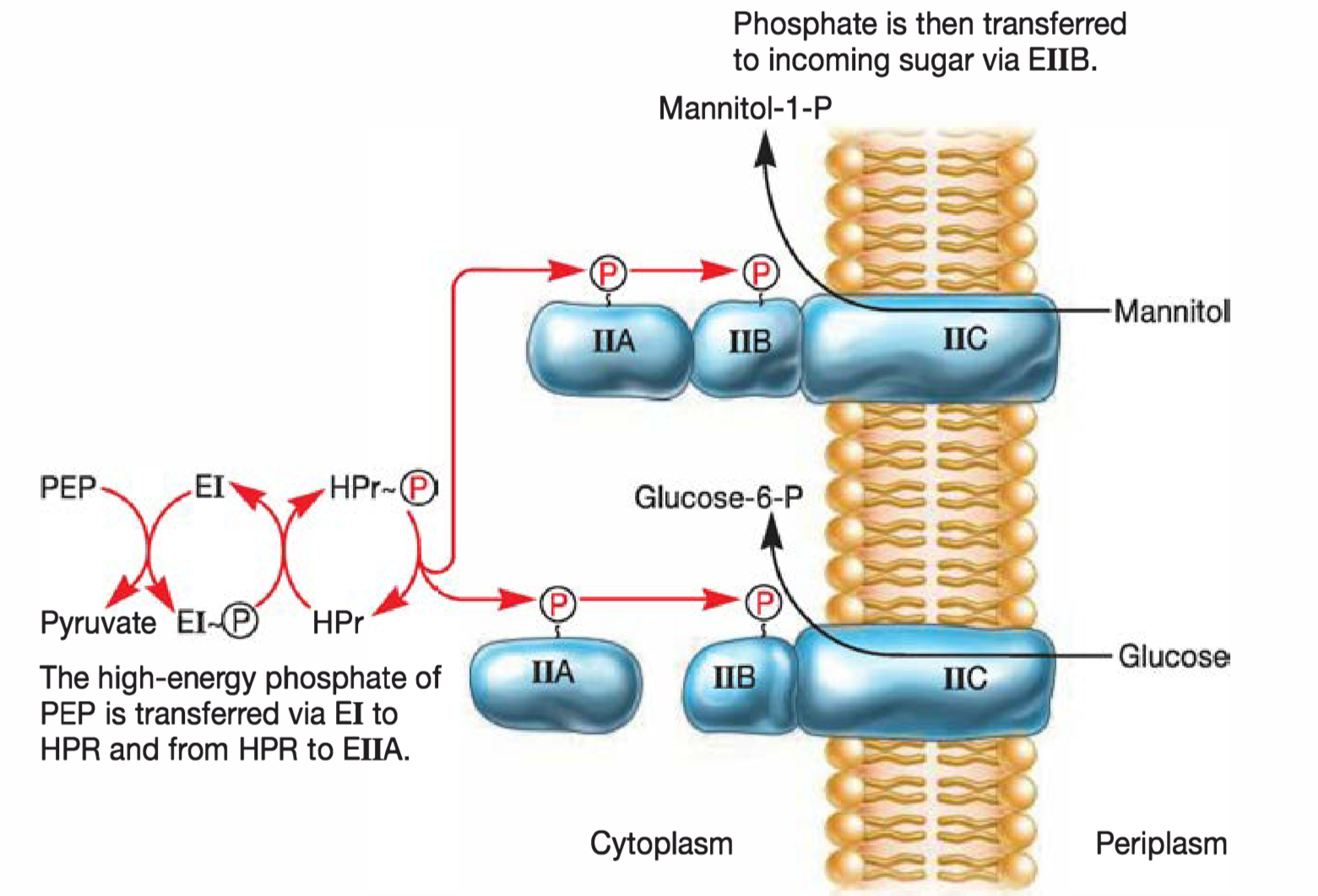
Group Translocation—Bacteria and Archaea
The distinguishing characteristic of group translocation is that a molecule is chemically modified as it is brought into the cell.
Energy dependent transport that chemically modifies molecule as it is brought into cell
Best known translocation system is phosphoenolpyruvate(PEP): sugar phosphotransferase system (PTS)
5. Ion uptake
以铁离子为例。
Ferric iron is very insoluble so uptake is difficult
Microorganisms secrete siderophores to aid uptake
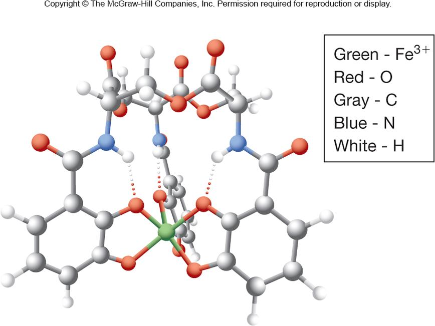
siderophores(铁载体),许多细菌通过分泌siderophores使得其与铁离子结合,再通过ABC transporters进入细胞
Bacterial Cell Walls
1. Functions
- Maintains the shape of the bacterium (Almost all bacteria have one)
- Helps protect cell from osmotic lysis
- Helps protect from toxic materials
- May contribute to pathogenicity
2. Peptidoglycan 肽聚糖(murein,胞壁质)
Meshlike polymer of identical subunits forming long strands
two alternating sugars:
A-acetylglucosamine (NAG)
N- acetylmuramic acid (NAM)
alternating D- and L-amino acids
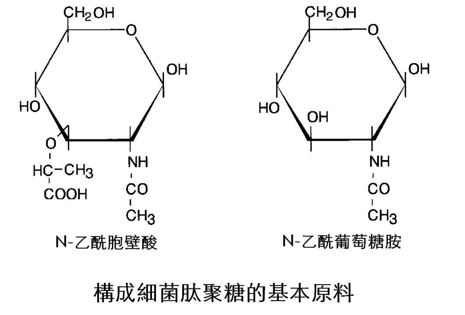
肽聚糖,存在于真细菌中的革兰氏阳性菌和革兰氏阴性菌的细胞壁中。肽聚糖的骨架是由两种糖衍生物:N-乙酰葡糖胺(N-acetylglucosamine,GlcNAc,NAG)和N-乙酰胞壁酸(N-acetylmuramic,MurNAc,NAM)交替相连而形成的多糖链,这些链相互交联形成肽聚糖。
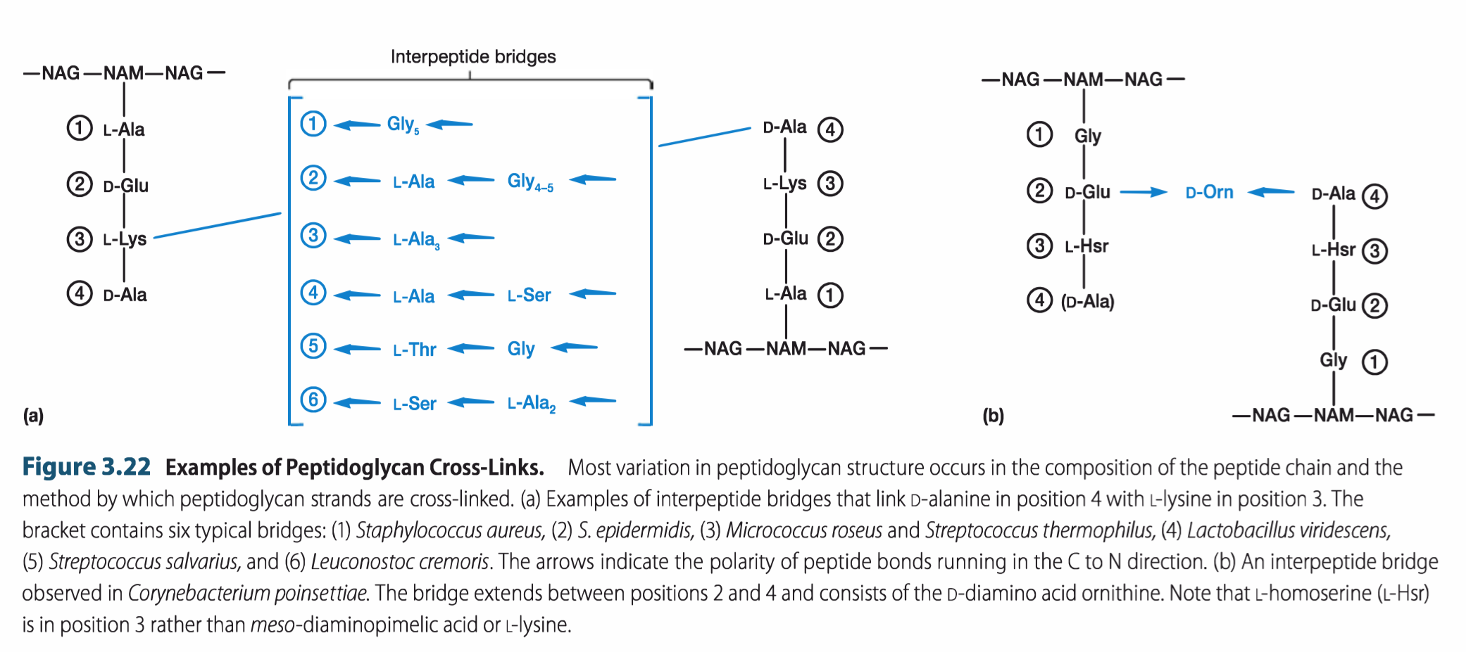
Strands Are Crosslinked
Peptidoglycan chains are crosslinked by peptides for strength
- interbridges may form
- peptidoglycan sacs 一 interconnected networks
- various structures occur
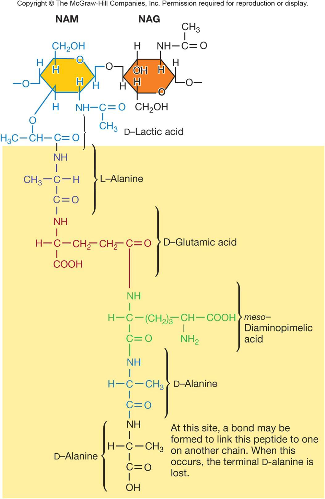
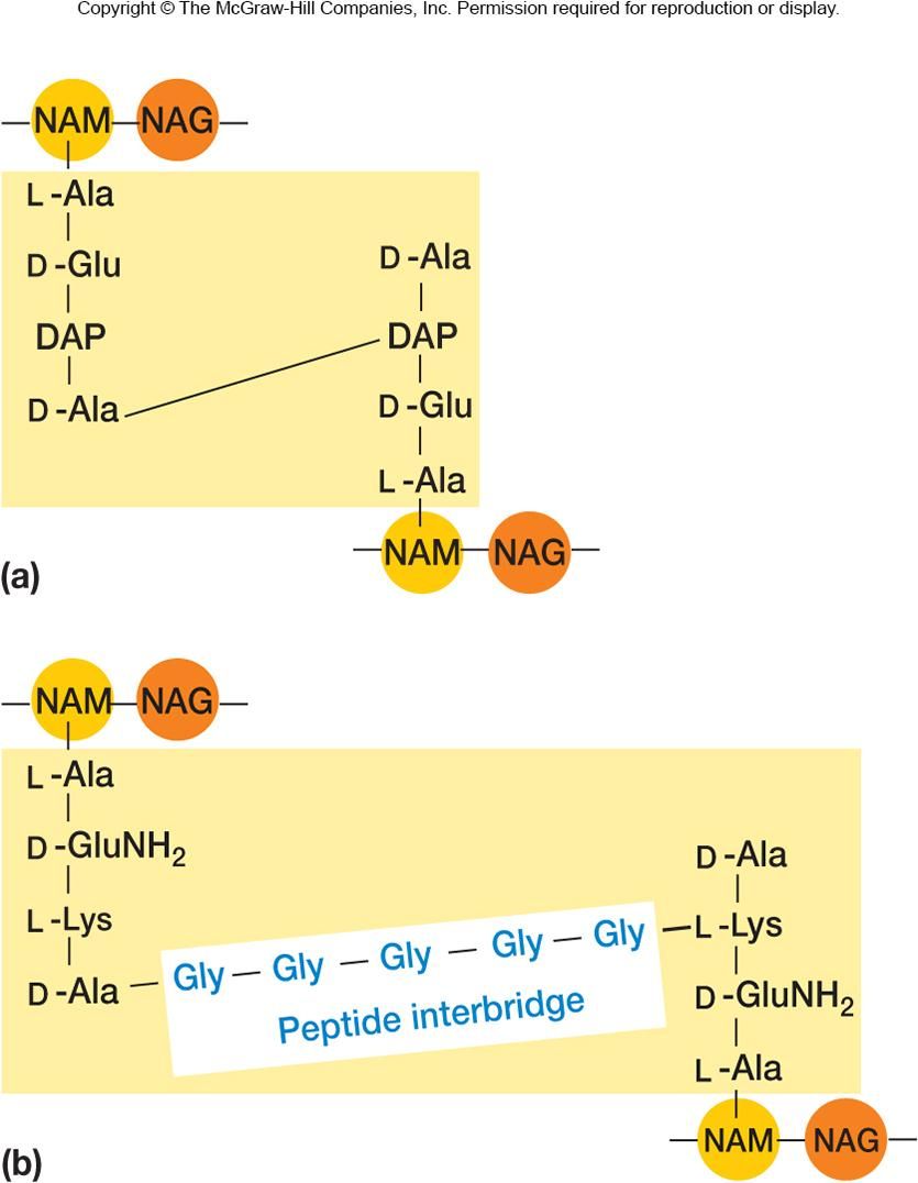
The peptidoglycan sacculus is formed by linking the sugars of the peptidoglycan subunits together to form a strand; the strands are then cross-linked to each other by covalent bonds formed between the peptides extending from each strand.
Peptidoglycan strands have a helical shape
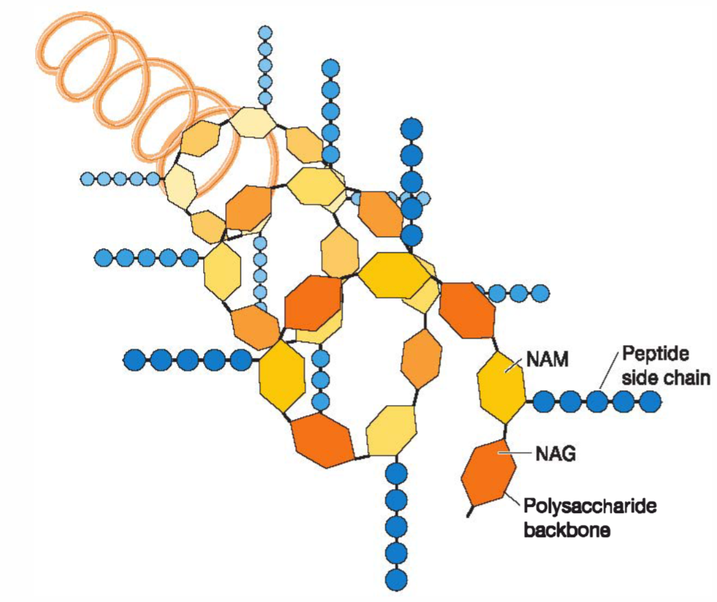
The strand is helical, and the peptides extend out from the backbone in different directions.
- Direct:Many bacteria cross-link the strands by connecting the carboxyl group of the D-alanine at position 4 directly to the amino group of diaminopimelic acid (position 3) of the other strand (the position 5o-alanine is removed as the cross-link is formed).
- Indirect:Other bacteria use a peptide interbridge instead (figure 3.19). With or without an interbridge, cross-link results in one dense, interconnected network of peptidoglycan strands
3. Gram-Positive Cell Walls
结构
Composed primarily of peptidoglycan
May also contain teichoic acids (negatively charged)
- help maintain cell envelope
- protect from environmental substances
- may bind to host cells
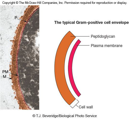
Most bacteria that stain Gram positive belong to the phyla Firmicutes and Actinobacteria, and most of these bacteria have thick cell walls composed of peptidoglycan and large amounts of other polymers such as teichoic acids(磷壁酸,(negatively charged))
Functions
- help maintain cell envelope
- protect from environmental substances
- may bind to host cells
Periplasmic Space of Gram + Bacteria
Lies between plasma membrane and cell wall and is smaller than that of Gram-negative bacteria
Periplasm has relatively few proteins
Enzymes secreted by Gram-positive bacteria are called exoenzymes
- aid in degradation of large nutrients
4. Gram-Negative Cell Walls
More complex than Gram-positive.
Consist of a thin layer of peptidoglycan surrounded by an outer membrane
Outer membrane composed of lipids, lipoproteins, and lipopolysaccharide (LPS)
No teichoic acids
结构
One of the most striking differences is the paucity of peptidoglycan. The peptidoglycan layer is very thin (2 to 7 nm, depending on the bacterium) and sits within the periplasmic space.
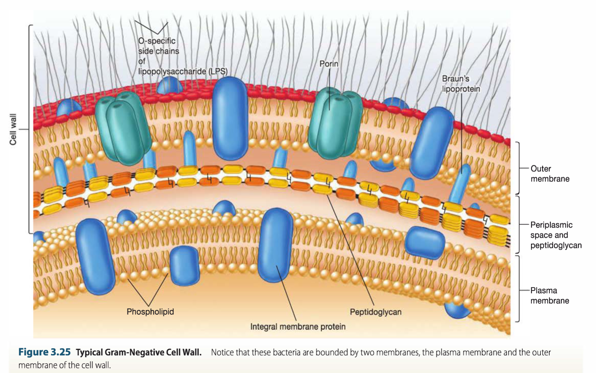
The outer membrane lies outside the thin peptidoglycan layer. It is linked to the cell by Braun’s lipoprotein(胞壁质脂蛋白), the most abundant protein in the outer membrane
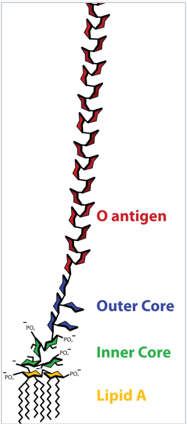
Lipopolysaccharides (LPSs,脂多糖). These large, complex molecules contain both lipid and carbohydrate, and consist of three parts: (1) lipid A, (2) the core polysaccharide, and (3) the O side chain(O antigen).
Peptidoglycan is -5-10% of cell wall weight
Periplasmic space differs from that in Gram-positive cells
may constitute 20-40% of cell volume
many enzymes present in periplasm
- hydrolytic enzymes, transport proteins and other proteins
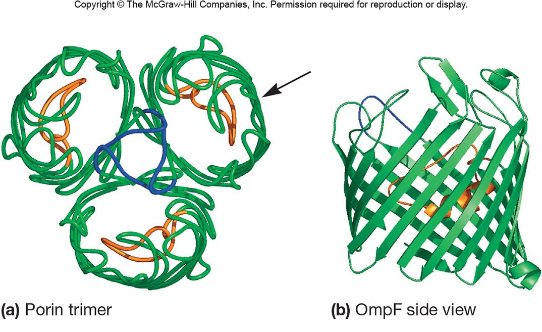
Gram-Negative Outer Membrane Permeability
More permeable than plasma membrane due to presence of porin proteins and transporter proteins
- porin proteins form channels to let small molecules (600-700 daltons) pass
5. Functions of LPS
Consists of three parts
- lipid A
- core polysaccharide
- O side chain (O antigen)
Lipid A embedded in outer membrane
Core polysaccharide, O side chain extend out from the cell
Importance of LPS
- contributes to negative charge on cell surface
- helps stabilize outer membrane structure
- may contribute to attachment to surfaces and biofilm formation
- creates a permeability barrier
- protection from host defenses (O antigen)
- can act as an endotoxin (lipid A)
It contributes to the negative charge on the bacterial surface because the core polysaccharide usually contains charged sugars and phosphate.
因为LPS负电荷较强,所以与磷壁酸一样,具有吸附Mg^2+^,Ga^2+^在细胞表面,提高其在细胞表面浓度的作用
It helps stabilize outer membrane structure because lipid A is a major constituent of the exterior leaflet of the outer membrane.
It helps create a permeability barrier. The geometry of LPS and interactions between neighboring LPS molecules are thought to restrict the entry of bile salts, antibiotics, and other toxic substances that might kill or injure the bacterium. 较小的分子可以透过,像溶菌酶、抗生素等较大的分子无法通过。
LPS also plays a role in protecting pathogenic bacteria from host defenses. 许多细菌可以快速改变它们的O antigen,从而阻碍宿主的免疫反应对其造成伤害。
The lipid A portion of LPS can act as a toxin and is called endotoxin. 脂质A是G^—^细菌致病物质内毒素的基础。
6. Mechanism of Gram Stain Reaction
shrinkage of the pores of peptidoglycan layer of Gram-positive cells
- constriction prevents loss of crystal violet during decolorization step
thinner peptidoglycan layer and larger pores of Gram-negative bacteria does not prevent loss of crystal violet
第一步初染剂(Primary Stain):使用结晶紫(crystal Violet)染色,染色部位主要为染细胞壁上的肽聚糖。
第二步媒染剂(Mordant):加入复方碘溶液或称革兰氏碘液(Gram’s Iodine)后,两者会形成不溶性的结晶紫-碘(CV-I)复合体。细菌呈紫黑色。第二步是关键,它的目的是分辨细菌的革兰氏染色特性。若为革兰氏阳性菌,此复合体系结合于细胞壁之镁、核糖核酸(Magnesium Ribonucletic Acid),形成镁-核糖核酸-结晶紫-碘(Mg-RNA-CV-I)复合体,不易脱去,染剂也就固定下来了。
第三步脱色剂(Decoloring Agent):使用酒精(95%)脱色,试剂具脂溶剂(Lipid Solvent)和蛋白脱水剂(Protein Dehydrating Agent)之双重功能。此步骤对革兰氏阳性细菌无效,因为其细胞壁上厚厚的细胞壁(成分为肽聚糖)层次多,交联致密,故遇脱水时网孔缩小,再加上不含类脂,使得乙醇不会溶出缝隙,因此阻隔了结晶紫-碘复合体的外渗,仍然呈紫色只能留在细菌体内。
革兰氏阴性菌细胞壁层薄,外膜类脂含量高、肽聚糖层薄和交联度差(Gram-negative cell walls is very thin, not as highly cross linked, and has larger pores),在遇脱色剂时,以类脂为主的外膜迅速溶解,薄而松散的肽聚糖网不能阻挡复合体溶出,因此会被脱色而成为无色。
第四步复染剂(Counterstain):为了对比,在此之后会加入番红(Safranin)进行复染,革兰氏阴性菌成红色,而革兰氏阳性菌仍保有原来的紫色。
7. Evidence of Protective Nature of the Cell Wall
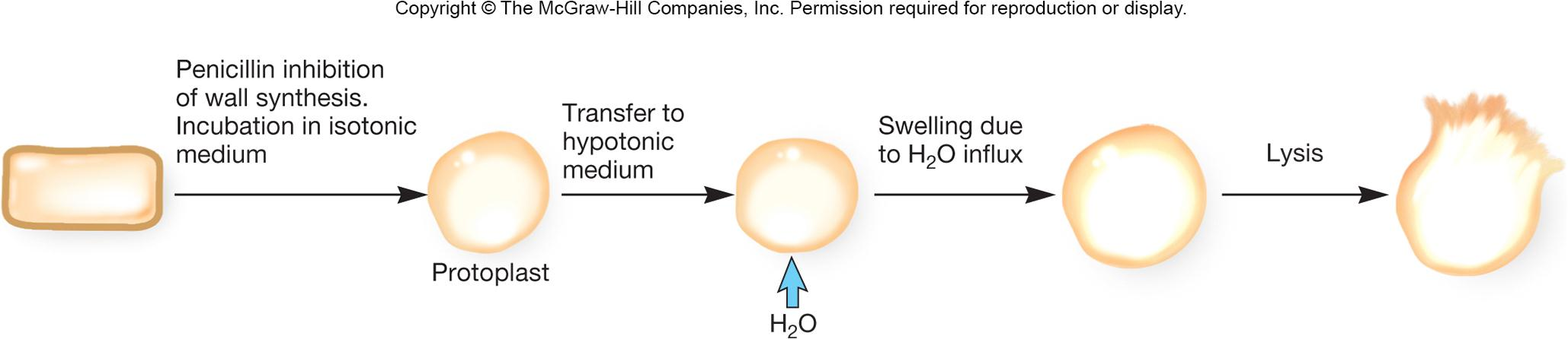
lysozyme breaks the bond between N-acetyl glucosamine and N-acetylmuramic acid
penicillin inhibits peptidoglycan synthesis
if cells are treated with either of the above they will lyse if they are in a hypotonic solution
Cell Envelope Layers Outside the Cell Wall
Components Outside of the Cell Wall
Outermost layer in the cell envelope
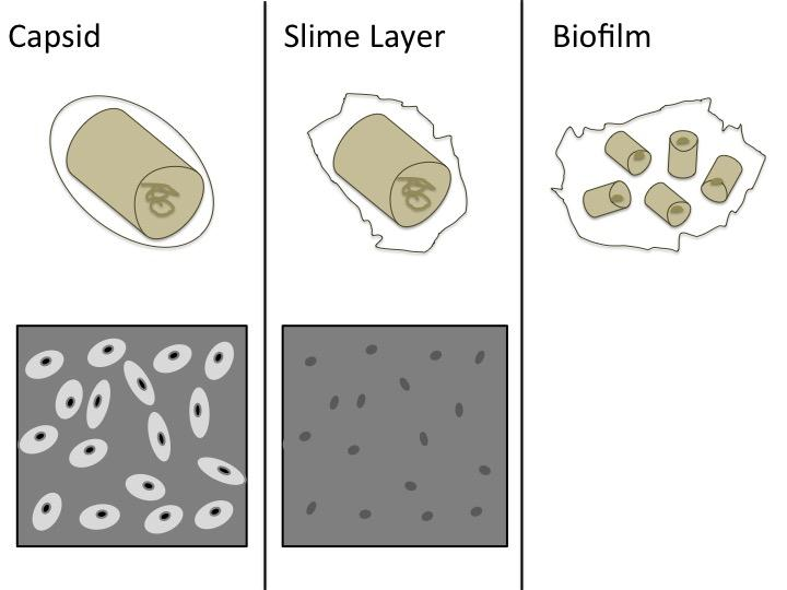
Glycocalyx
- capsules, slime layers and biofilms
S layers
1. Capsules(荚膜) and Slime Layers(黏液层)
Glycocalyx(糖萼,多糖包被):糖胶层是对由细菌、上皮细胞,或其他细胞分泌的,覆盖在细胞表层的粘稠物的统称。Capsules and Slime Layers are all glycocalyx.
Capsules
Usually composed of polysaccharides
Well organized and not easily removed from cell
Visible in light microscope
Protective advantages
- resistant to phagocytosis
- protect from desiccation
- exclude viruses and detergents
Capsules are most often composed of polysaccharides, but some are constructed of other materials.
(Bacillus anthracis(炭疽杆菌) has a proteinaceous capsule composed of poly D-glutamic acid.)
Functions:
- They help pathogenic bacteria resist phagocytosis(吞噬作用) by host phagocytes. why
- Capsules can also protect against desiccation because they contain a great deal of water. They exclude viruses and most hydrophobic toxic materials such as detergents(洗涤剂)
Slime Layers
similar to capsules except diffuse, unorganized and easily removed
slime may aid in motility
黏液层(slime layer)为一种细菌的特殊构造,它是围绕细菌细胞的一层很容易除去(比如通过离心的方法),而且无规则的物质。黏液层的主要化学成分为外多糖、糖蛋白,以及糖脂。
黏液层的功能是保护细菌细胞免受来自环境的伤害,诸如抗菌素和干燥的环境。黏液层亦可以使细菌附着到光滑的表面上,比如义肢类医疗设备和导管。黏液层能保护菌落免被含氯制剂、含碘制剂等化学灭菌剂杀死,使得高压蒸汽灭菌法或沸水冲洗成为唯一的去污染方法。
黏液层与S-Layers不同
Biofilm
生物薄膜(英语:biofilm),也称作“生物膜”或“菌膜”,是一些微生物细胞由自身产生的胞外多聚物基质(主要为多糖)所包围而形成,且附著在浸有液体的惰性或生物表面的,具有结构的群落。
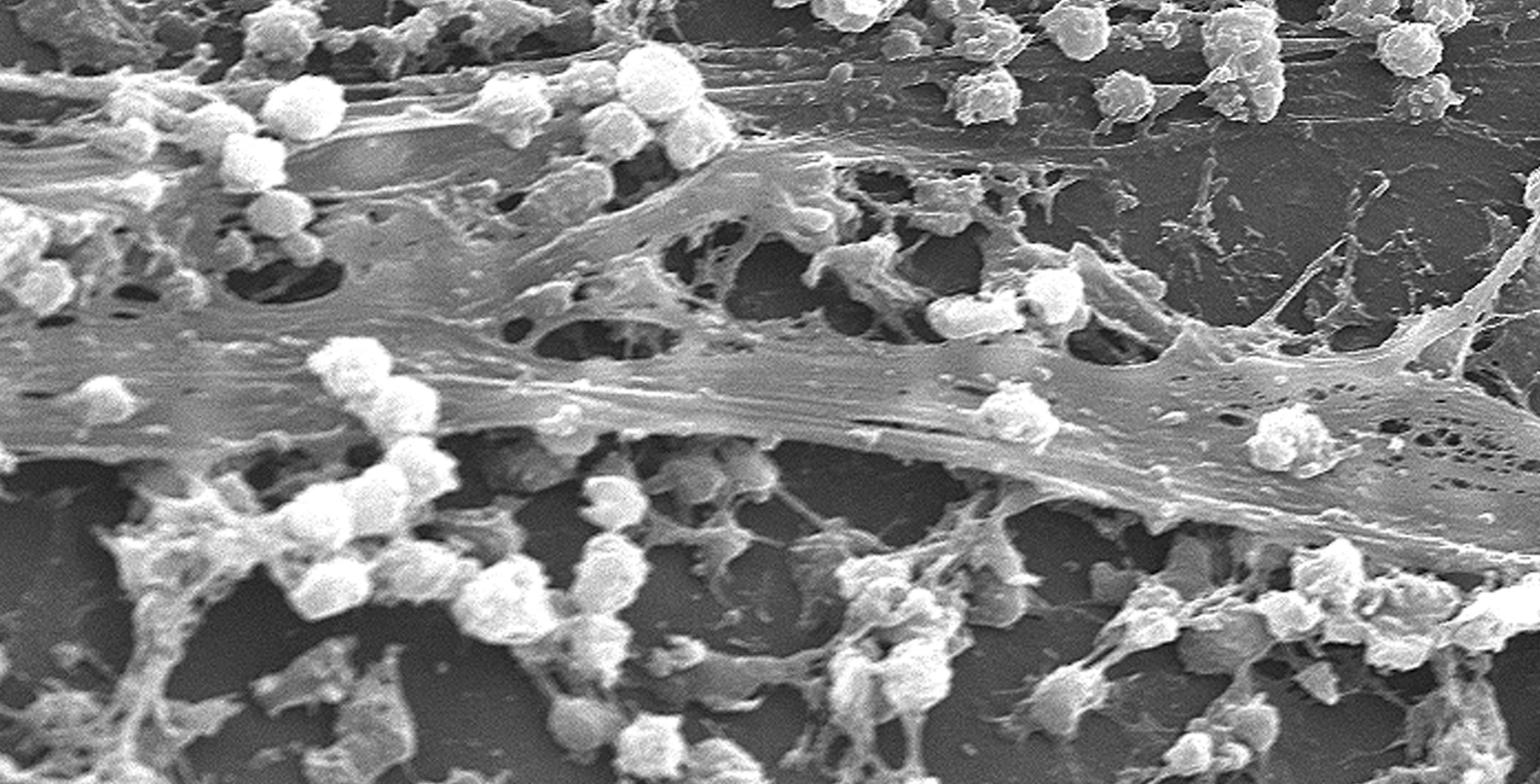
2. S-Layers (Surface Layers)
Regularly structured layers of protein or glycoprotein that self-assemble
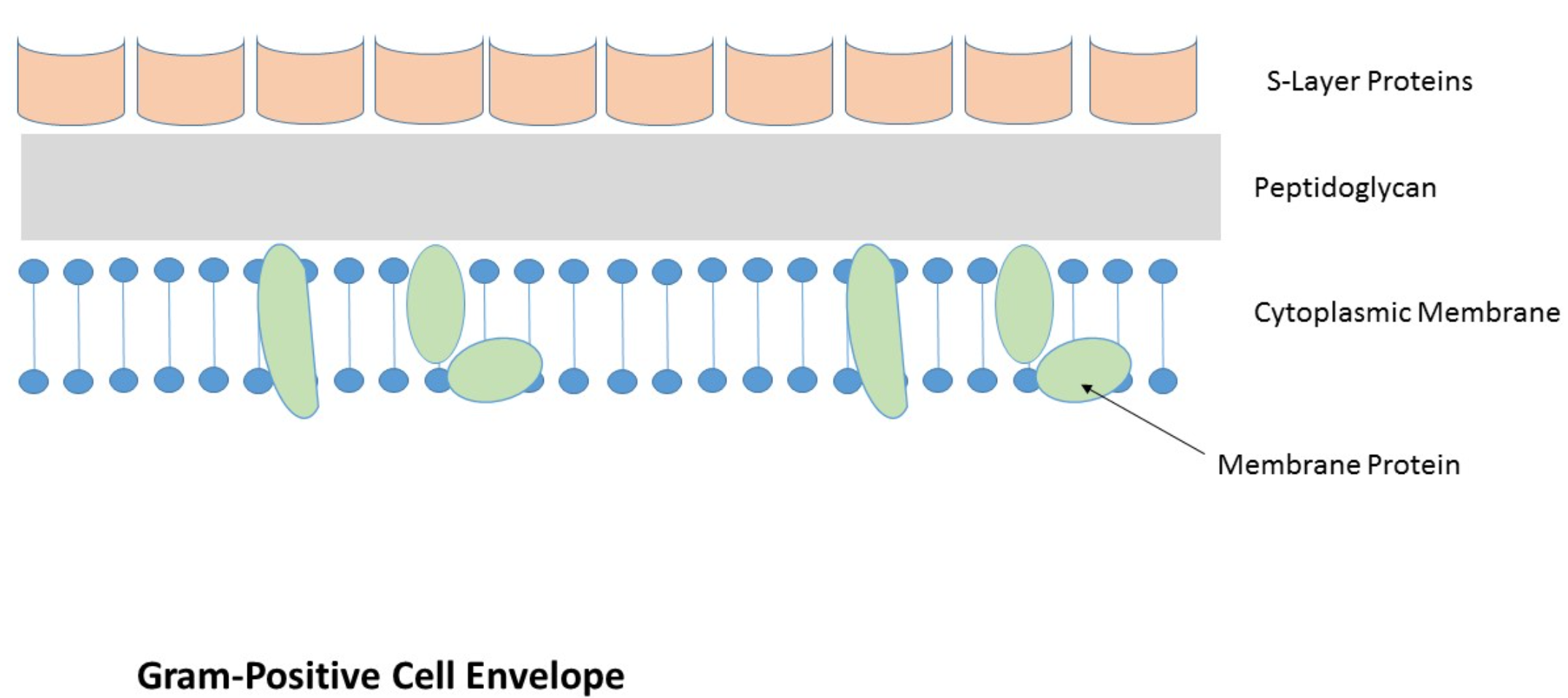
S层(S-layer, surface layer)是古菌和细菌细胞包被的一部分。S层由糖蛋白,蛋白质和铺成的单分子组成。S层通过分子的自组装形成,会覆盖整个细胞表面。
Functions:
- Protect from ion and pH fluctuations, osmotic stress, enzymes, or predatory bacteria
- Maintain the shape and envelope rigidity of some cells
- Promote cell adhesion to surfaces
- protect some bacterial pathogens against host defenses
- Potential use in nanotechnology
- S layer spontaneously associates
Bacterial Cytoplasm
**Protoplast: **The plasma membrane and everything within
**cytoplasm: **The material bounded by the membrane.
- Cytoskeleton
- Intracytoplasmic membranes
- Inclusions
- Ribosomes
- Nucleoid and plasmids
1. Bacterial Cytoskeleton
Homologs of all 3 eukaryotic cytoskeletal elements have been identified in bacteria
Functions are similar as in eukaryotes
Three elements
Eukaryotes possess three cytoskeletal elements: actin actin filaments(微丝), microtubules(微管), and intermediate filaments(中间纤维,中间丝).
Actin filaments are made from actin, and microtubules are made from tubulin. Inter mediate filaments are composed of a mixture of one or more members of different classes of proteins.
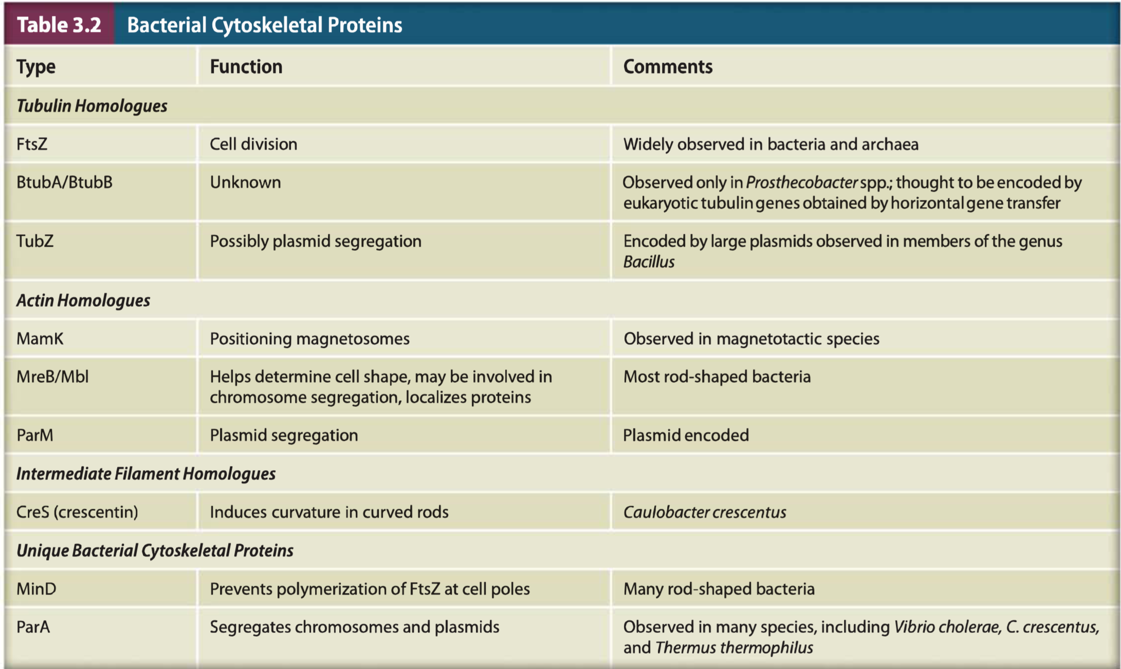
(1) Examples
FtsZ 一 many bacteria
- forms ring during septum formation in cell division
MreB - many rods
- maintains shape by positioning peptidoglycan synthesis machinery
CreS 一 rare, maintains curve shape
Three model cells
The cytoskeletons of Escherichia coli(大肠杆菌), Bacillus subtilis(枯草杆菌), and Caulobacter crescentus(新月柄杆菌) are the best studied and are the focus of our discussion. These three organisms are important bacterial model systems for several reasons.
2. Intracytoplasmic Membranes 胞质内膜
在一些细菌细胞中,同样可以观察到内膜结构。
Plasma membrane infoldings
- observed in many photosynthetic bacteria
- observed in many bacteria with high respiratory activity
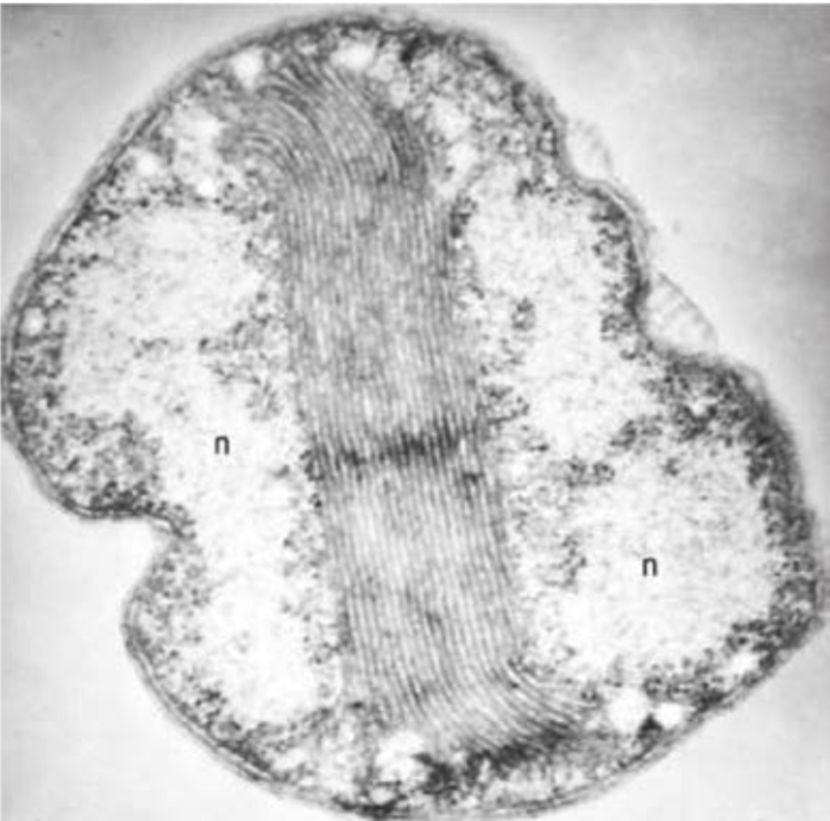
These can be extensive and complex in photosynthetic bacteria and in bacteria with very high respiratory activity, such as the nitrifying bacteria(硝化细菌).
The internal membranes of the photosynthetic cyanobacteria(蓝细菌) are called thylakoids(类囊体) and are analogous(同功的) to the thylakoids of chloroplasts.
However, these internal membranes differ from the plasma membrane by being enriched for proteins and other
molecules involved in energy conservation.
The membrane-bound organelle called the anammoxosome(厌氧氨氧化体) is observed in some members of the phylum Planctomycetes and deserves additional comment
3. Inclusions(细胞质内含物)
Granules of organic or inorganic material that are stockpiled by the cell for future use
Some are enclosed by a single-layered membrane
- membranes vary in composition
- some made of proteins; others contain lipids
Inclusions can take the form of granules(粒料), crystals(晶体), or globules(水珠,小球体); some are amorphous(无固定形状的).
Storage Inclusions
Storage of nutrients, metabolic end products, energy, building blocks
Glycogen storage
Carbon storage
- poly-β-hydroxybutyrate (PHB)
Phosphate - Polyphosphate (Volutin, 异染质)
Amino acids - cyanophycin granules
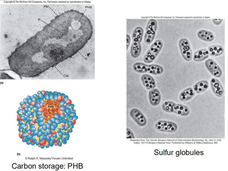
Cells have a wide variety of storage inclusions. Many are formed when one nutrient is in ready supply but another nutrient is not. Some store end products of metabolic processes.
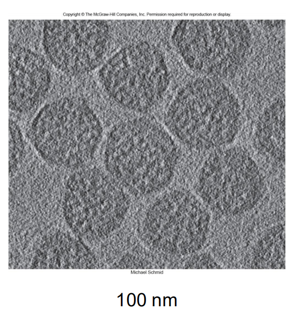
Microcompartments
Not bound by membranes but compartmentalized for a specific function
Carboxysomes - $CO_{2}$ fixing bacteria
- contain the enzyme used for $CO_{2}$ fixation
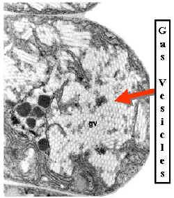
Other Inclusions
(1) Gas vacuoles
found in aquatic, photosynthetic bacteria and archaea
provide buoyancy in gas vesicles
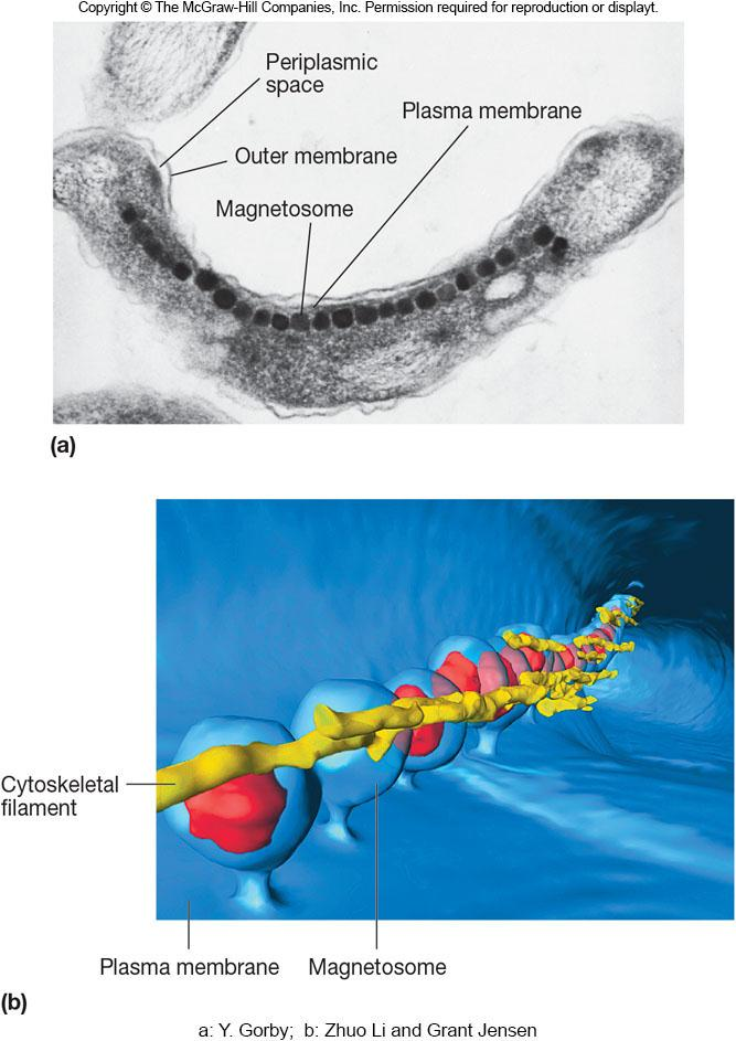
(2) Magnetosomes 磁小体
found in aquatic bacteria
magnetite particles for orientation in Earth’s magnetic field
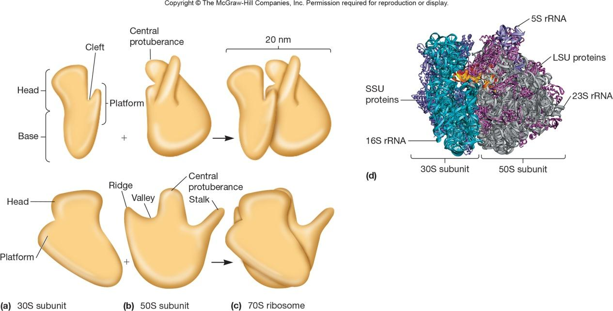
4. Ribosomes
Complex protein/RNA structures
bacterial and archaea ribosome = 70S
eukaryotic (80S) S = Svedburg unit
Bacterial ribosomal RNA
- 16S in small subunit
- 23S and 5S in large subunit
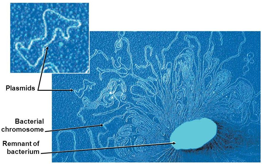
5. The Nucleoid
Usually not membrane bound (few exceptions)
Usually 1 closed circular, double-stranded DNA molecule
Supercoiling and nucleoid proteins (different from histones) aid in folding
6. Plasmids
Extrachromosomal DNA
- found in bacteria, archaea, some fungi
- usually small, closed circular DNA molecules
Exist and replicate independently of chromosome
- inherited during cell division
Contain few genes that are non-essential
External Structures
Extend beyond the cell envelope in bacteria
- pili and fimbriae
- flagella
Function in cell movement, protection, attachment to surfaces, horizontal gene transfer
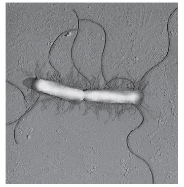
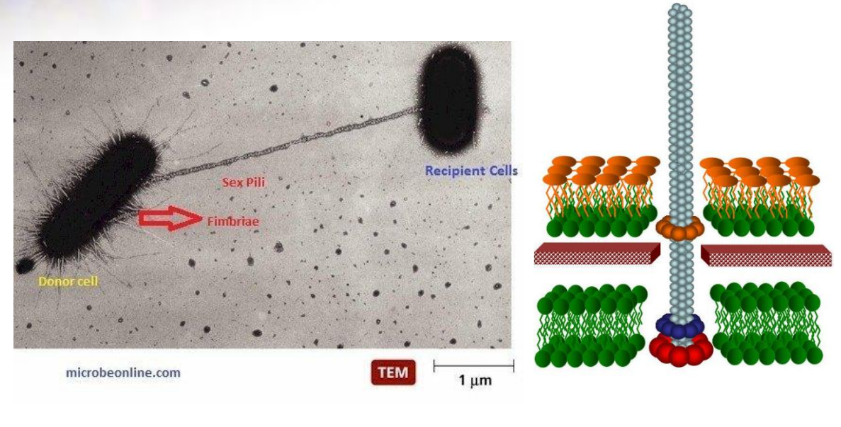
Fimbriae(菌毛) and Pili
Fimbriae (s., fimbria)
- short, thin, hairlike, proteinaceous appendages (up to 1,000/cell)
- can mediate attachment to surfaces, motility, DNA uptake
Sex pili (s., pilus)
- longer, thicker, and less numerous (1-10/cell)
- required for conjugation
- gene transfer
Pili are primarily composed of oligomeric pilin proteins, which arrange helically to form a hollow cylinder.
2. Flagella 鞭毛
Threadlike, locomotor appendages extending outward from plasma membrane and cell wall
Functions
motility and swarming behavior
attachment to surfaces
may be virulence factors
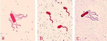
Patterns of Flagella Distribution
Monotrichous(单鞭毛) 一 one flagellum
Polar flagellum(极性鞭毛) 一 flagellum at end of cell
Amphitrichous(双鞭毛的) 一 one flagellum at each end of cell
Lophotrichous (丛鞭毛的,偏端丛毛的)一 cluster of flagella at one or both ends
Peritrichous(周生鞭毛的) 一 spread over entire surface of cell
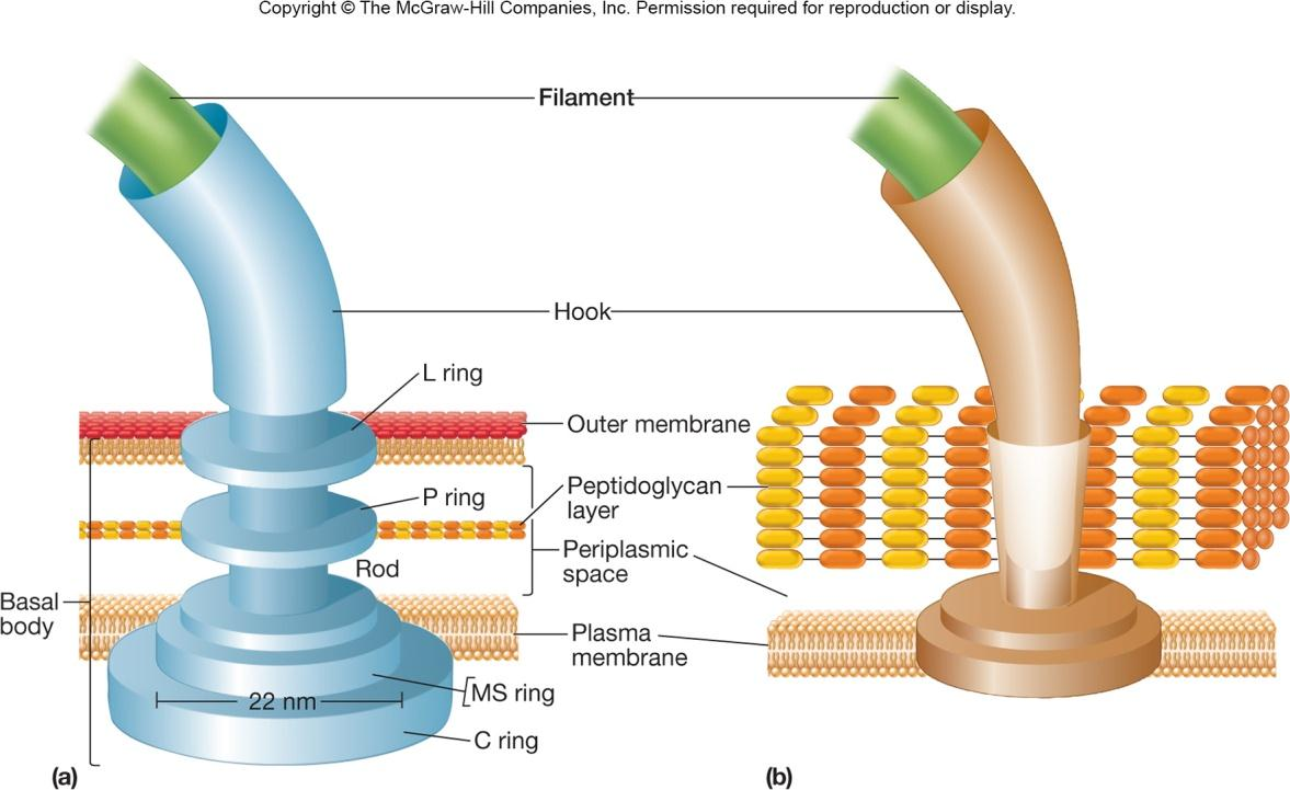
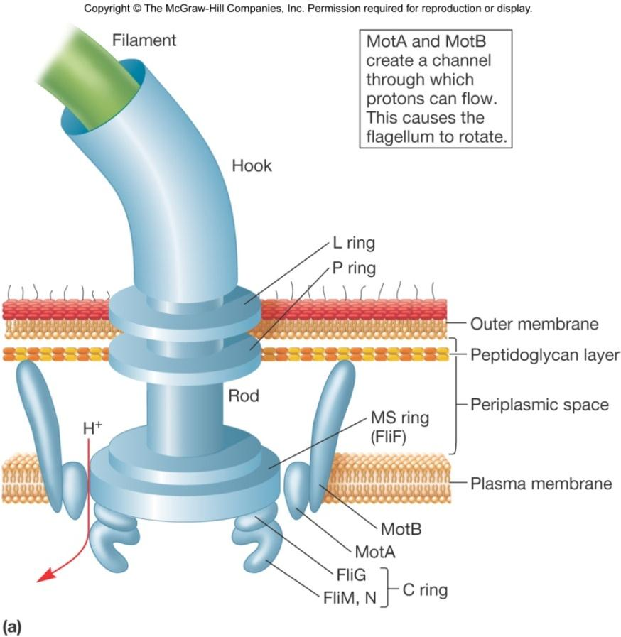
Three Parts of Flagella
Filament
- extends from cell surface to the tip
- hollow, rigid cylinder of flagellin protein
Hook
- links filament to basal body
Basal body
- series of rinqs that drive flaqellar motor
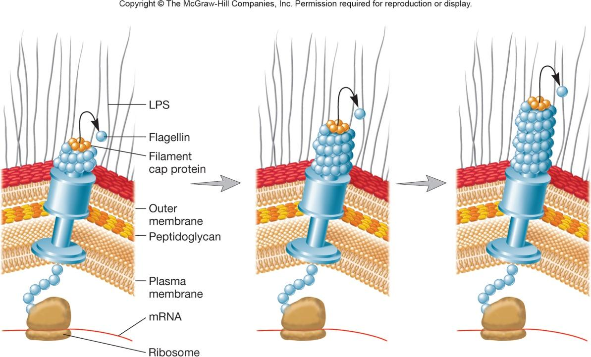
3. Flagellar Synthesis
complex process involving many genes/gene products
new flagellin molecules transported through the hollow filament using Type Ill-like secretion system
filament subunits self-assemble with help of filament cap at tip, not base
三、Bacterial Motility and Chemotaxis
Motility
Flagellar movement 鞭毛运动
Spirochete motility 螺旋运动
Twitching motility 蹭行运动
Gliding motility 滑行运动
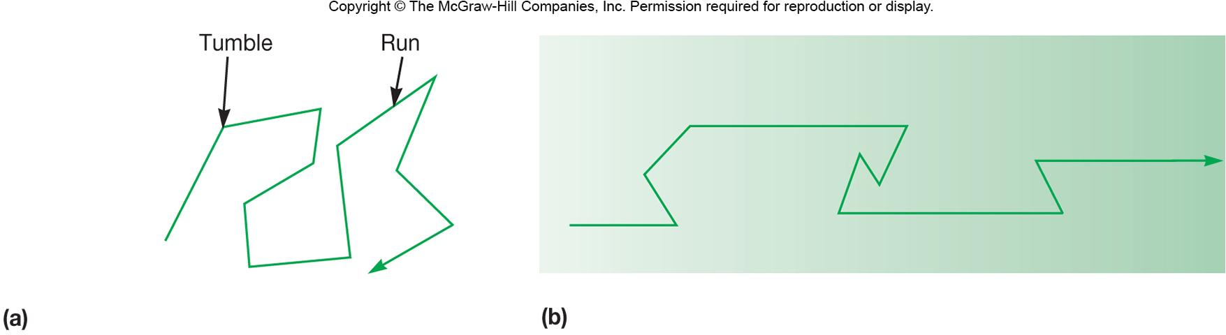
1. Bacterial Flagellar Movement
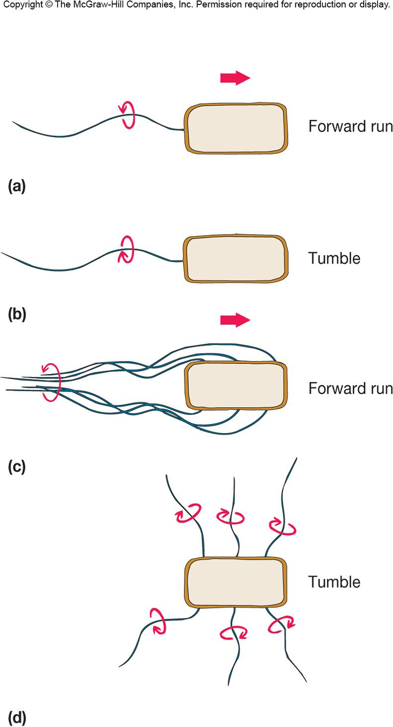
Flagellum rotates like a propeller
- very rapid rotation up to 1100 revolutions/sec
- in general, counterclockwise (CCW) rotation causes forward motion (run)
- in general, clockwise rotation (CW) disrupts run causing cell to stop and tumble
Mechanism
Flagellum is 2 part motor
Rotor
- C (FliG protein) ring and MS ring turn and interact with stator
Stator Mot A and Mot B proteins
form channel through plasma membrane
protons move through Mot A and Mot B channels using energy of proton motive force
torque powers rotation of the basal body and filament
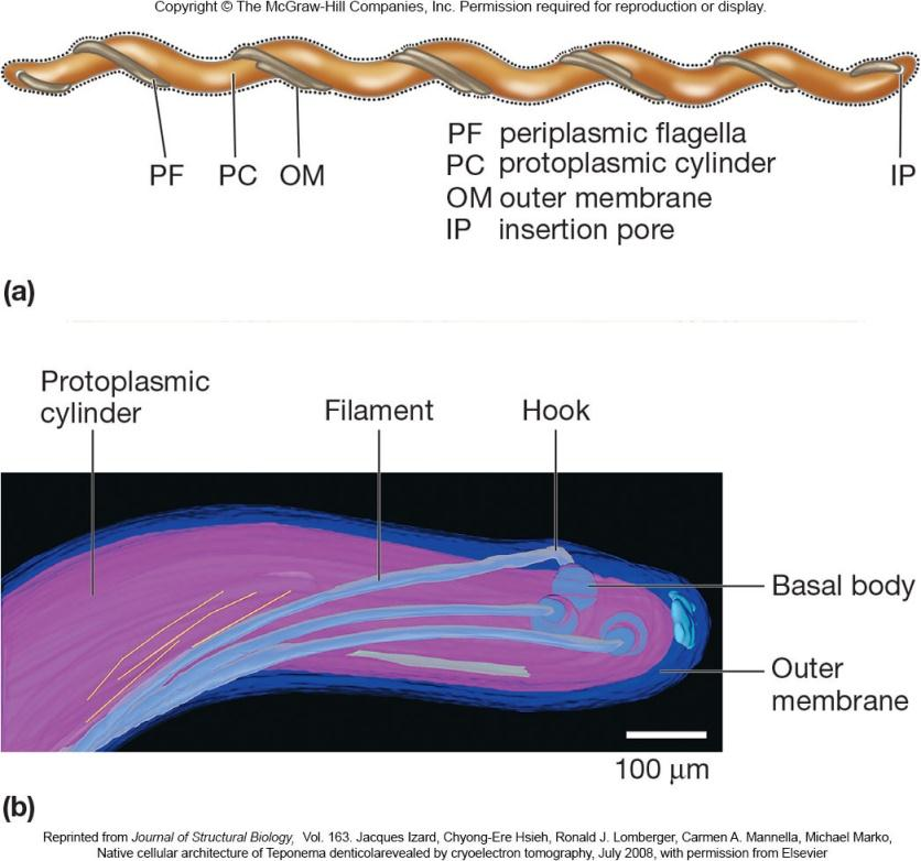
2. Spirochete Motility
Multiple flagella form axial fibril which winds around the cell
Corkscrew shape exhibits flexing and spinning movements
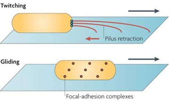
3. Twitching and Gliding Motility
Twitching
- pili at ends of cell
- short, intermittent(间歇的;断断续续的;间歇性), jerky motions
Gliding
- smooth movements
Chemotaxis
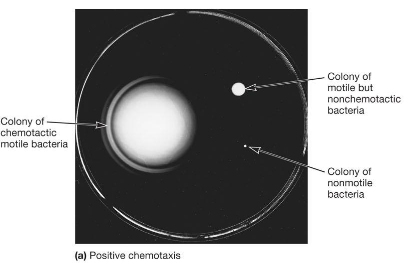
In presence of attractant (b) tumbling frequency is intermittently reduced and runs in direction of attractant are longer
Chemotaxis away from repellent involves similar but opposite responses
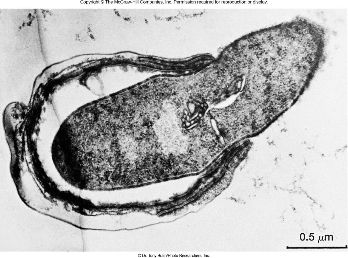
四、The Bacterial Endospore
Complex, dormant structure formed by some bacteria
Various locations within the cell
Resistant to numerous environmental conditions
- heat
- radiation
- chemicals
- desiccation
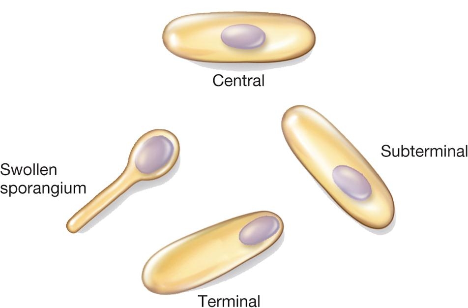
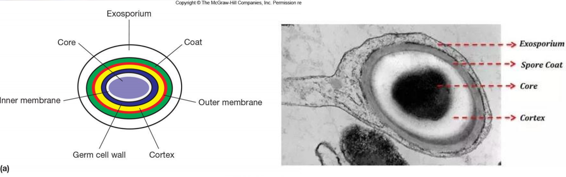
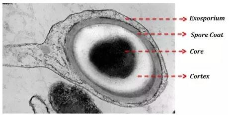
Endospore Structure
Spore surrounded by thin covering called exosporium
Thick layers of protein form the spore coat
Cortex, beneath the coat, thick peptidoglycan
Core has nucleoid and ribosomes
Reason of Endospore Resistance
Calcium (complexed with dipicolinic acid)
Small, acid-soluble, DNA-binding proteins (SASPs)
Dehydrated core
Spore coat and exosporium protect
Sporulation 孢子形成
Process of endospore formation
Complex multistage process
Occurs in a hours (up to 10 hours)
Normally commences when growth ceases because of lack of nutrients
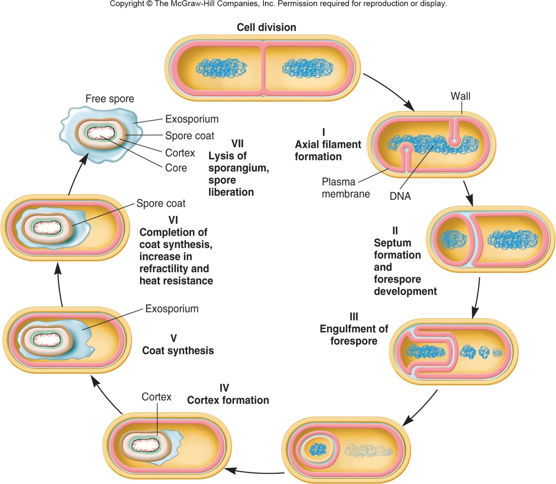
Formation of Vegetative Cell
Activation
- prepares spores for germination
- often results from treatments like heating
Germination
- environmental nutrients are detected
- spore swelling and rupture of absorption of spore coat
- increased metabolic activity
Outgrowth - emergence of vegetative cell