L9 Neurons
一、Neurons
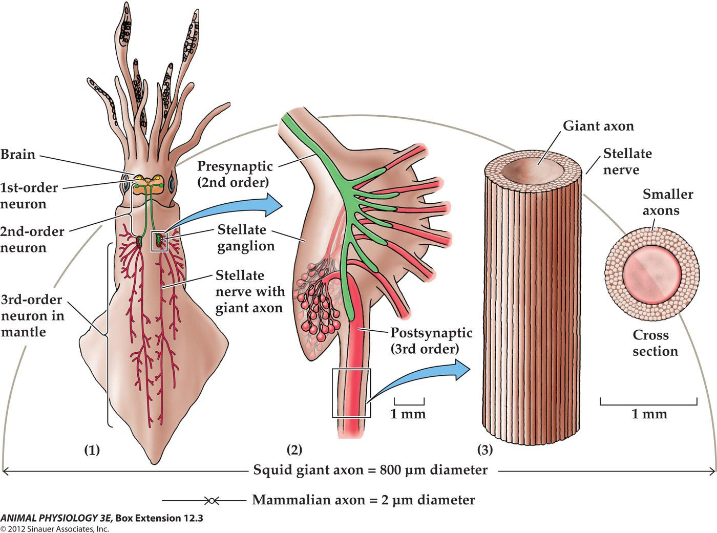
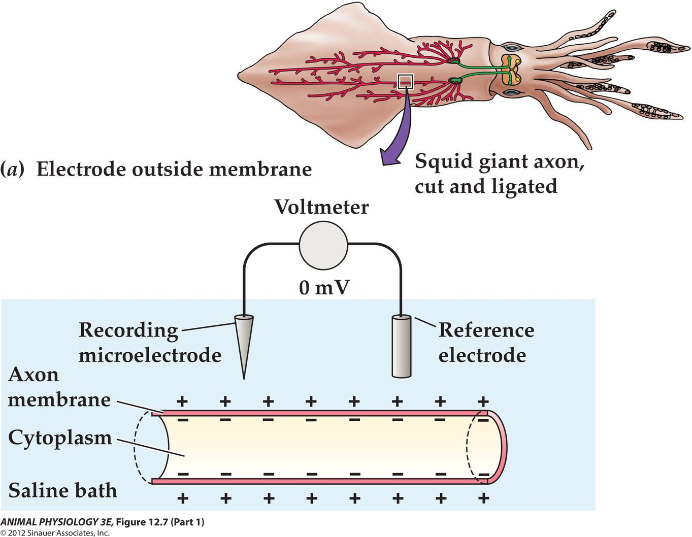
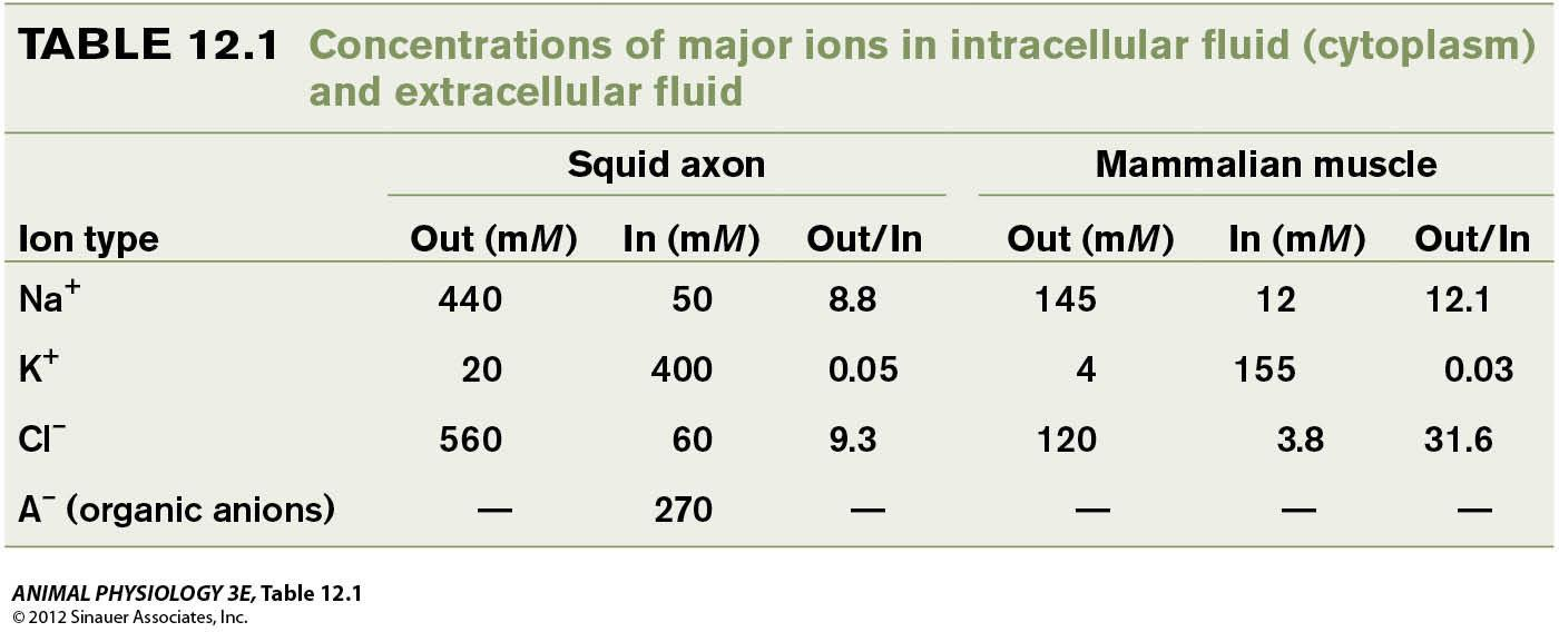
Squid giant axon:
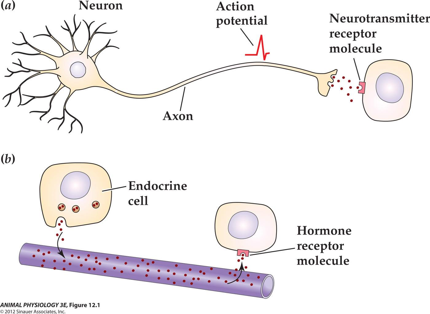
The physiology of control: neurons and endocrine cells compared.
Cellular integration refers to processes within cells;
Whole-animal integration refers to the selective combination of and processing of sensory, endocrine and central nervous system information in ways that promotes the harmonious functioning of the whole organism.
Types
Neuronal and hormonal signaling both convey information over long distances
Neurons have four functional regions that typically correspond to their four major structural regions
Functions
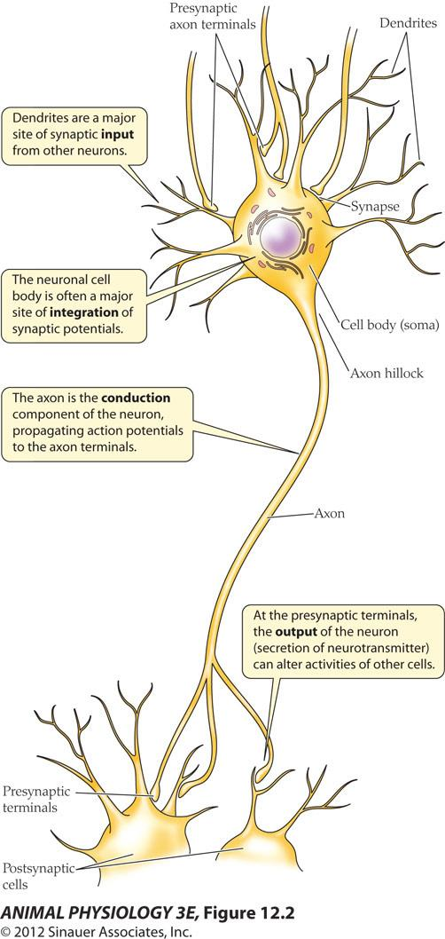
Neurons have four parts and four functions
- Dendrites – receives input
- Neuronal body – integration
- Axon – conduction
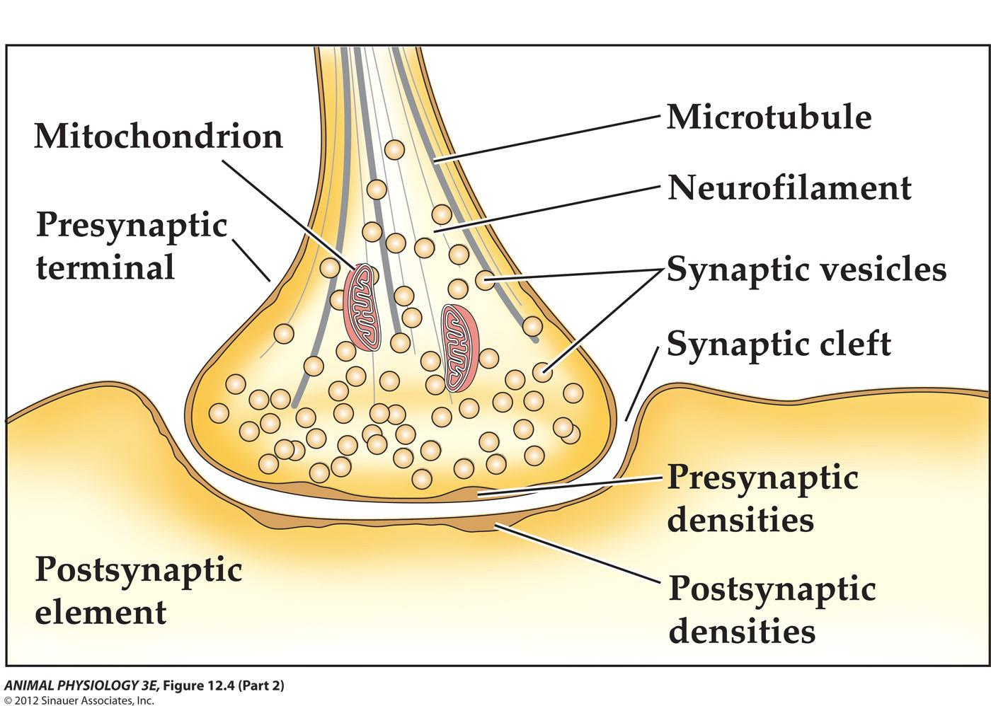
- Presynaptic terminal – output
Innervation – neurons form synaptic
Endings on a cell are said to innervate that cell
1. Neural Transmittion
Neurons transmit electrical signals to target cells
Central nervous system (CNS) - brain and spinal cord in vertebrates
Peripheral nervous system
Sensory (afferent)neurons –relay signals to integrative centers of the CNS
Motor (Efferent) neurons –relay control signals (instructions) from the CNS to target cells that are under nervous control, such as muscle cells and secretory cells.
Interneurons – neurons are entirely within the CNS
2. Endocrine cells broadcast hormones
In contrast to signals from neurons, signals from the endocrine system are broadly distributed throughout the animals body
Two essential features of the endocrine control: slow and broadcast – hormone is released in the blood and all cells are bathed in it.
Nervous system and endocrine systems control different processes:
- Neural communication is much finer both temporally and spatially than is possible for endocrine systems.
- Therefore, the nervous system controls the fine,rapid movements of discrete muscles
- The endocrine system typically controls more widespread.Prolonged activities such as metabolic changes.
- Nervous system and endocrine systems can exert control over each other as well as over other targets
3. Neurons are organized into functional circuits in nervous systems
The function of a nervous system depends on the wiring!
Take the next slide of cockroach’s jump reflex as an example.
- The neural circuit mediating the startle response in the cockroach Periplaneta americana
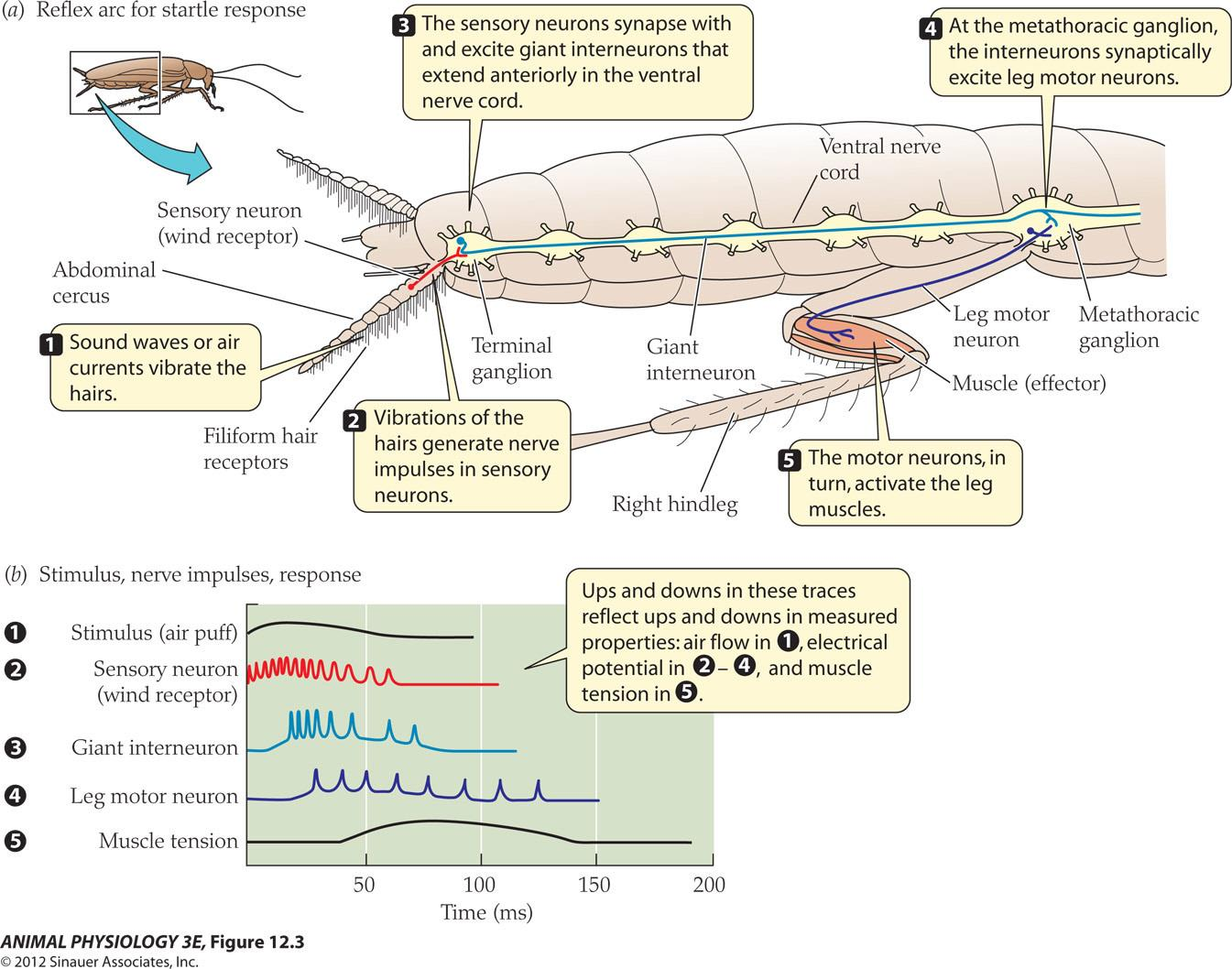
- This startle response takes less than 150 milliseconds
The cellular structure of neurons
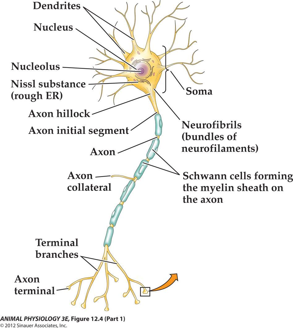
- An axon is single and long With constant diameter and few collateral branches
- Myelin sheath – not every axon is myelinated
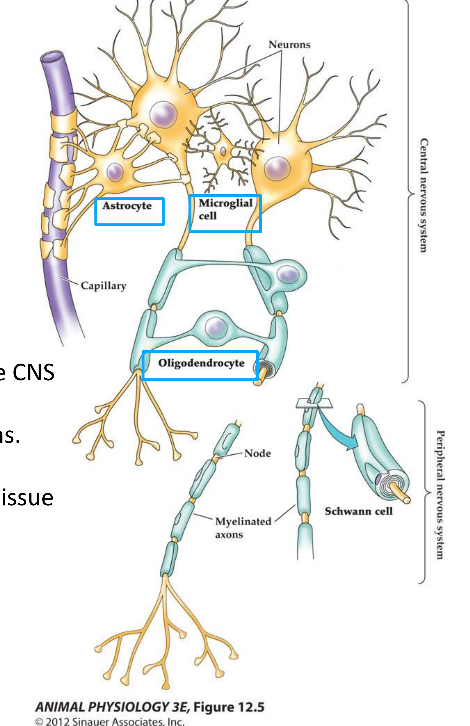
- Glial cells are estimated to make up half the volume of the mammalian brain and to outnumber neurons by ten to one.
- Schwann cells unsheathe PNS axons Oligodendrocytes are in the CNS.
- Astrocytes line the surface of the capillaries in the CNS To take up neurotransmitters from extracellular space and supply metabolic substances to neurons.
- Microglia mediates immune responses in neural tissue And may act as phagocytes in brain injury.
1. Changes in the membrane potential
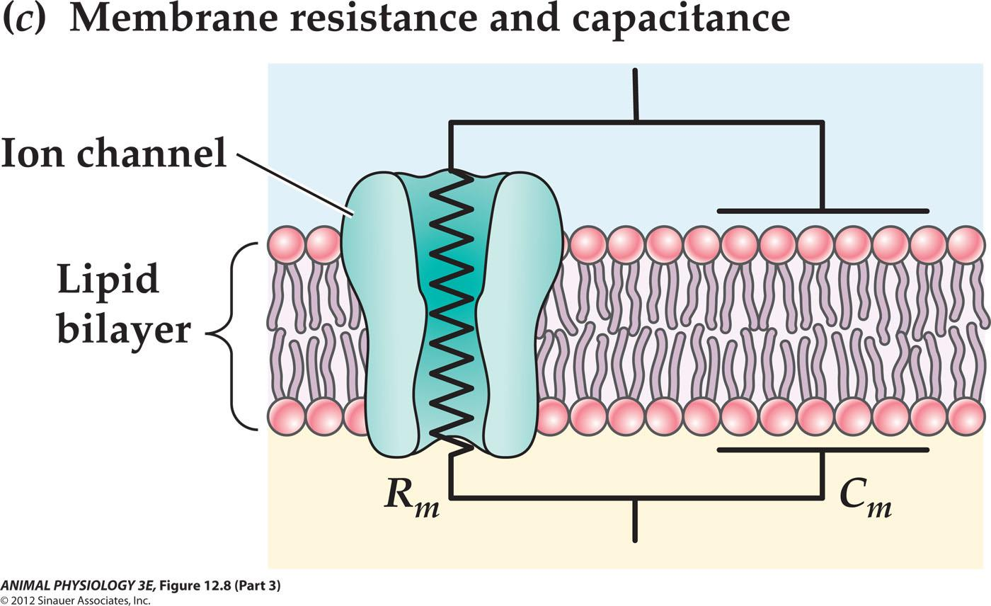
- The resistance corresponds to ion channels through which ions can flow
- The lipid bilayer corresponds to the dielectric layer of a capacitor that separates charges on its surface
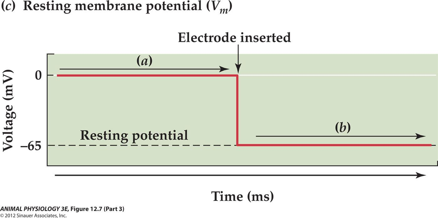
2. Recording the resting membrane potential of a squid giant axon
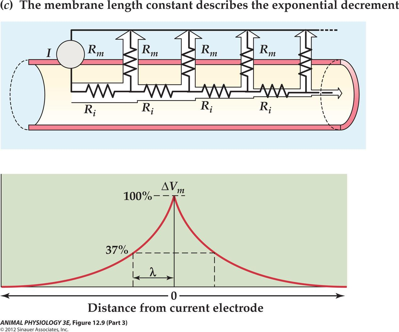
Changes in the membrane potential: the membrane time constant:
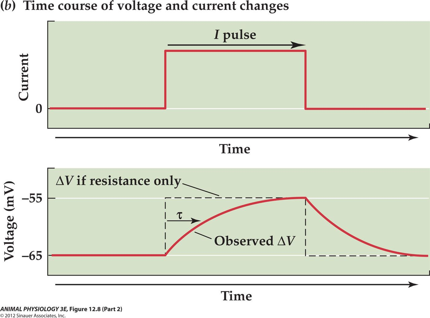
- The time constant tau is the time required for the observed ∆V to reach 63% of the maximum
- Ohm’s law:
- Graded potential ∆V=IR
- time constant tau = RC
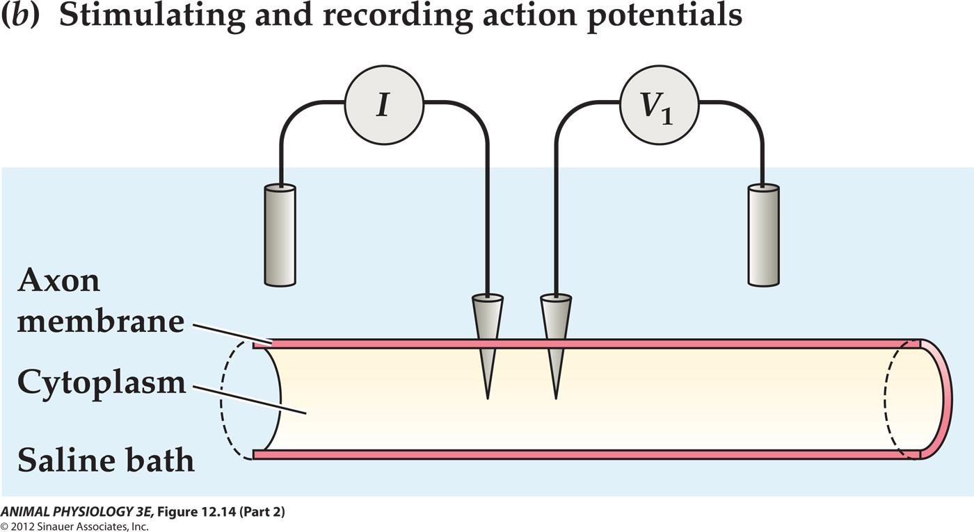
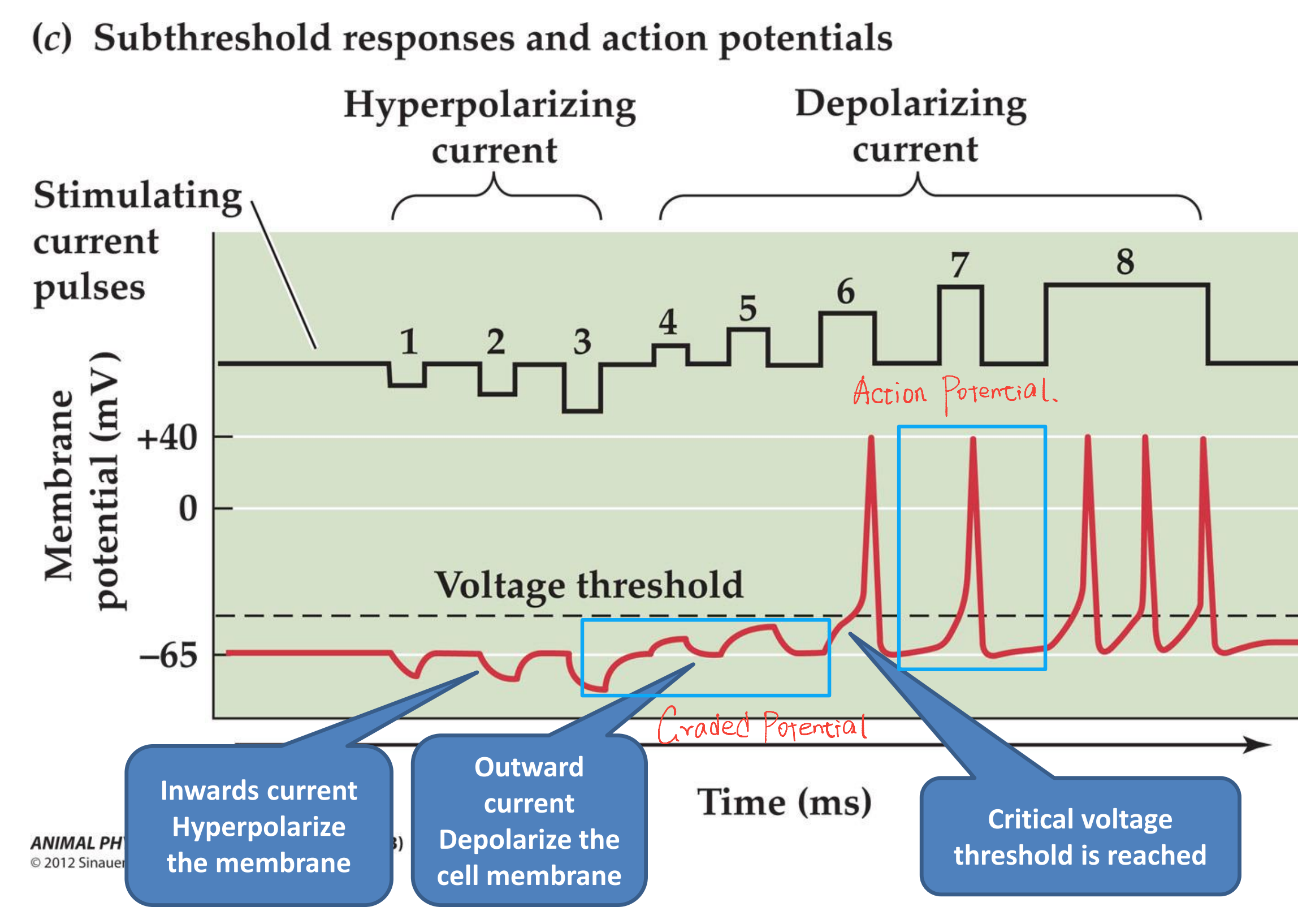
3. General features of action potentials
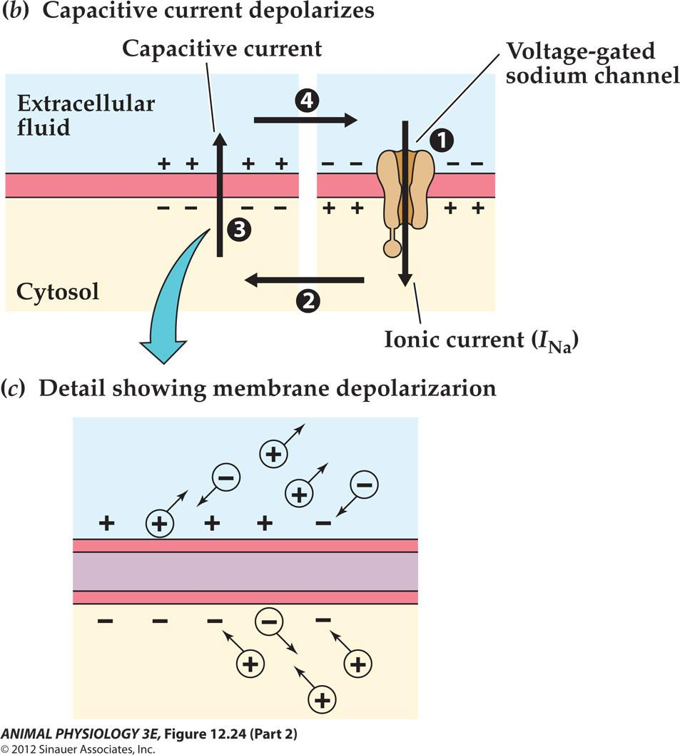
The Hodgkin cycle produces the rising phase of the action potential:
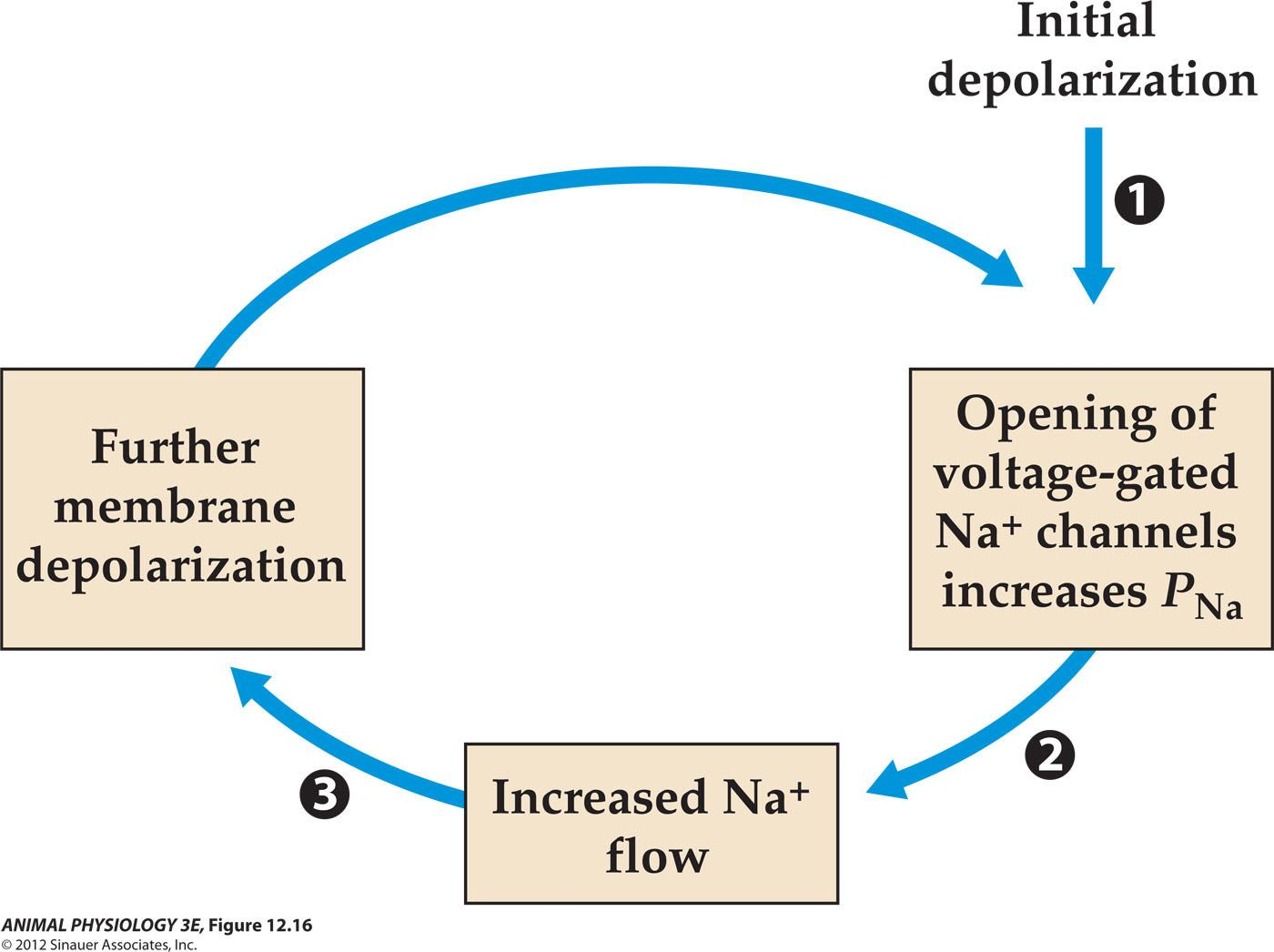
- Because most of the current flows through shorter paths of lower resistance, less and less current is available to change the membrane potential at greater distance from the source
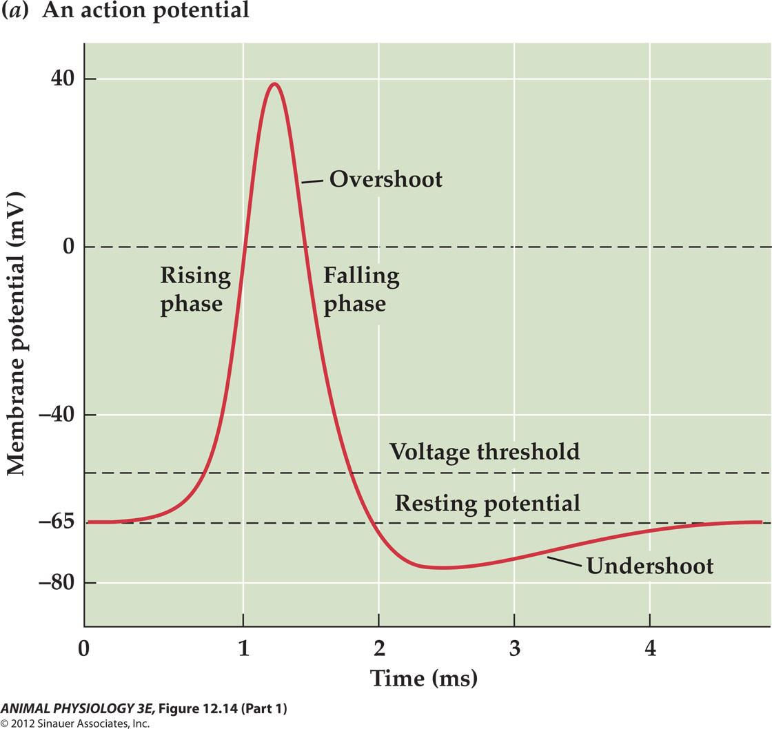
- Action potentials are voltage Dependent, ALL-or-NONE electrical signals
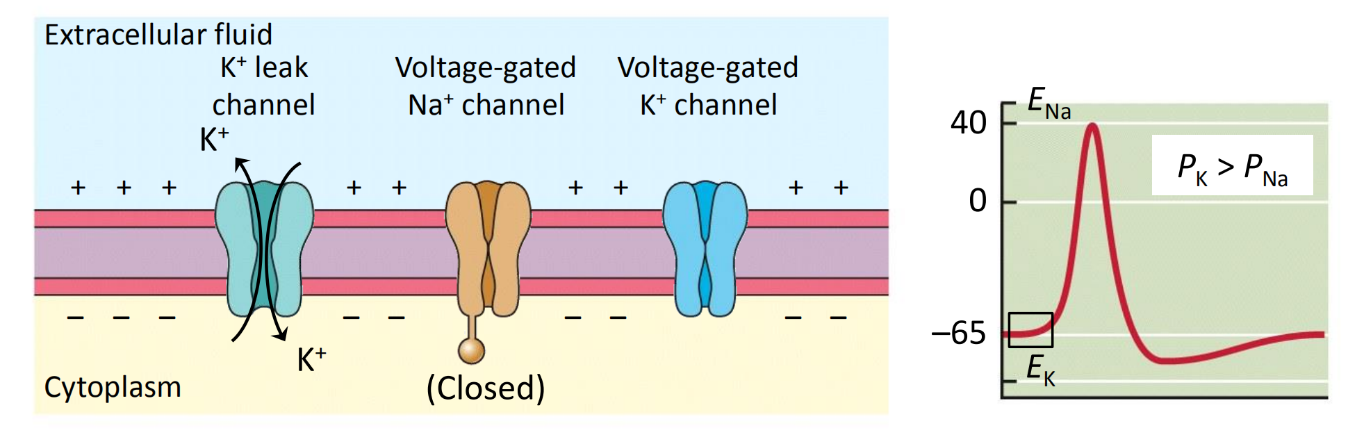
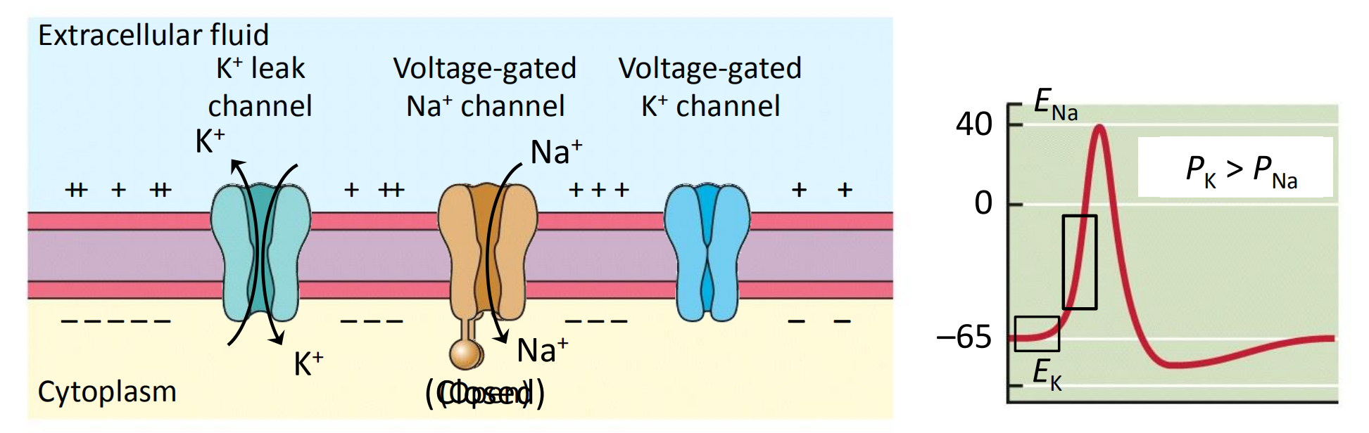
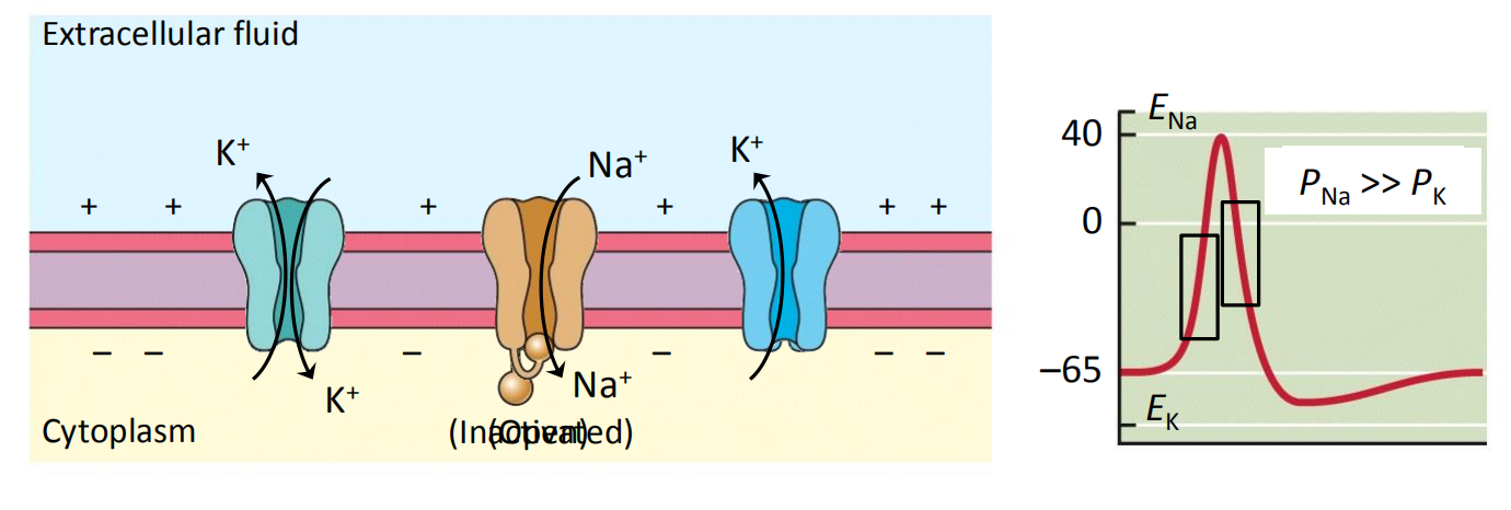
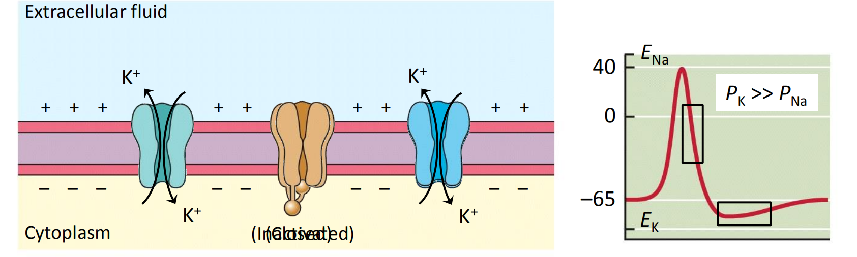
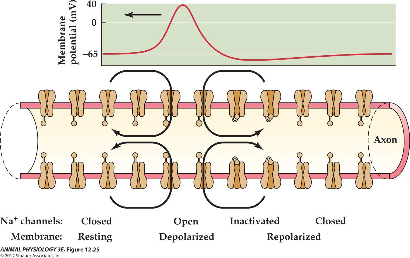
4. Membrane permeability changes that produce an action potential
- Resting membrane potential
- Rising phase
- Falling phase
- Recovery
Nonspiking neurons do not generate action potentials
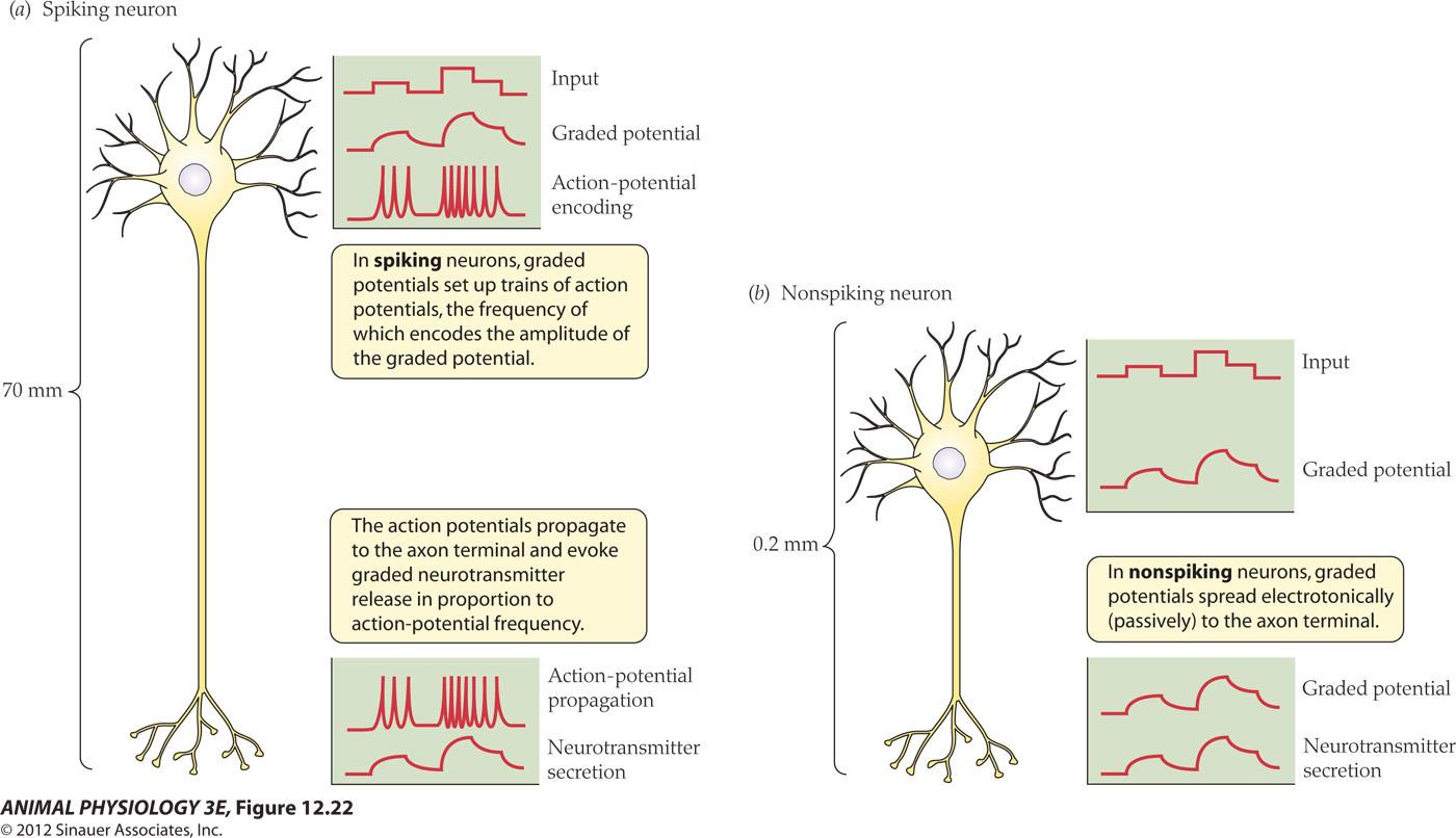
- 都产生了neurotransmitter release
- Nonspiking neurons: photoreceptors, Bipolar cells and horizontal cells of the vertebrate retina
二、The propagation of action potentials
Nondecremental propagation of the action potential is possible because the action potential at one location on an axon can itself initiate an action potential at a neighboring location and the induced action potential will have the all-or-none amplitude as the original action potential. By repeating this process, a signal can travel 1 m along an axon without any decrease in amplitude.
Membrane refractory periods prevent bidirectional propagation
Why don’t local currents initiate reverse-traveling impulses going the other direction?
- The membrane behind a traveling impulse is not re-excited by the local currents because a traveling impulse is still in its refractory period.
- Inactivation of Na channel prevents the channels from entering the Hodgkin cycle until the action potential is far enough away to minimize local depolarization (basis for absolute refractory period).
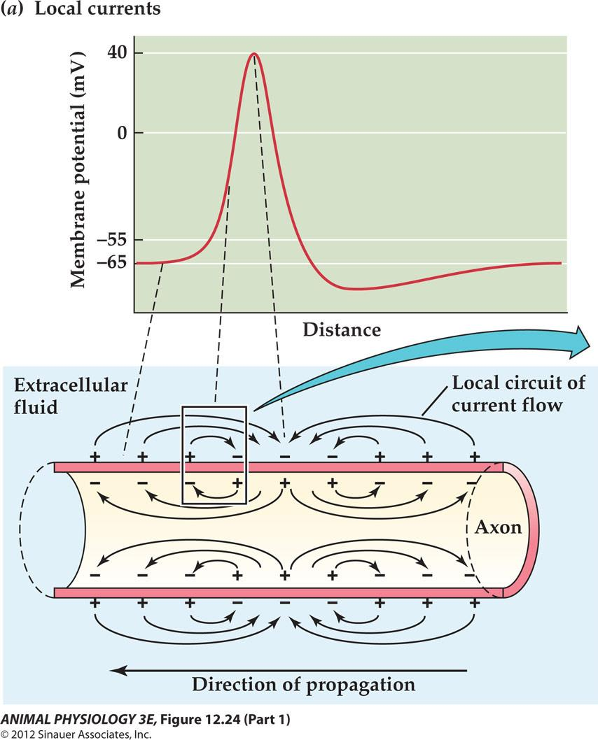
Inactivation of voltage-gated Na+ channels prevents reverse propagation of an action potential
The conduction velocity
The conduction velocity of an action potential depends on axon diameter, myelination, and temperature
- Large diameter axons tend to conduct action potentials more rapidly than small diameter axons
Myelination increases conduction velocity
- Myelinated axons represent huge evolutionary advantage as they allow very high conduction velocities with relatively small axon diameters.
- Wrapped with 200 or more concentric layers of glial membrane (Schwann cells for PNS and oligodendrocytes for CNS)
- The glial cytoplasm is excluded from between the glial membrane layers
- The insulating layer is called myelin, which stops at intervals of 1 mm or so. The gap is called node of Ranvier ( the only place allows ion flow across the axon membrane)
- Action potential only occurs at the Nodes of Ranvier in contrast to the continuous sweep of action potentials over an unmyelinated axon
1. Myelinated axons speed the propagation of an action potential
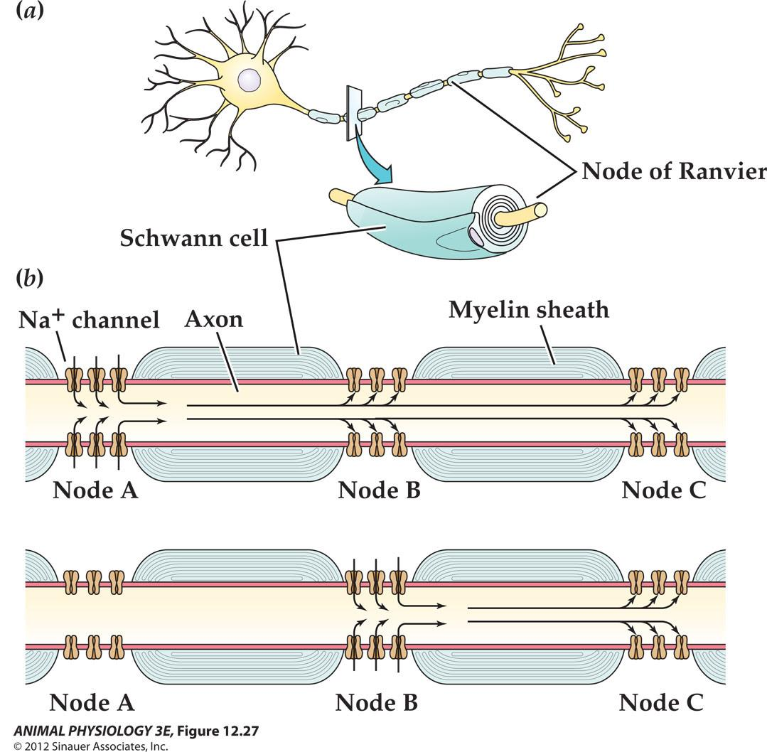
跳跃式传导 Saltatory conduction
- Action potential jumps from node to node
- Action Potential is imitated typically at the axon initial Segment.
- Myelinated membrane Resistance increased 1000-10000 fold over the Resistance at the node of Ranivier
2. The effect of myelin
To increase the membrane resistance of myelinated axon by 1000 fold!
To decrease membrane capacitance
- Capacitance is inversely proportional to the distance separating the charges on the plates of a capacitator, in this case, the distance between the axoplam and the extracellular fluid
- Myelin increases this distance in proportion to the number of glial membrane wrappings, so that capacitance is decreased about 1000-fold!
- time constant tau = RC
The increase in resistance (Rm) of myelinated region is offset by a decrease in Cm and membrane time constant is nearly unaffected.
Myelination greatly increased conduction velocity by increasing the axon length constant without increasing the time constant
3. Reduction in axon diameter
Myelination allows the same velocity to be achieved with a 40-fold reduction in diameter and a 1600-fold reduction in axon cross-sectional area and volume
- A frog myelinated axon 12 um in diameter has a conduction velocity of 25 m/s at 20 °C;
- An unmyelinated squid giant axon must be about 500 um in diameter to achieve the same 25 m/s velocity at 20 °C.
With the reduction in axon dimension, many more axons can be incorporated into a nervous system of a given size.
Spatial distribution of voltage-gated channels at the surface of a myelinated neuron
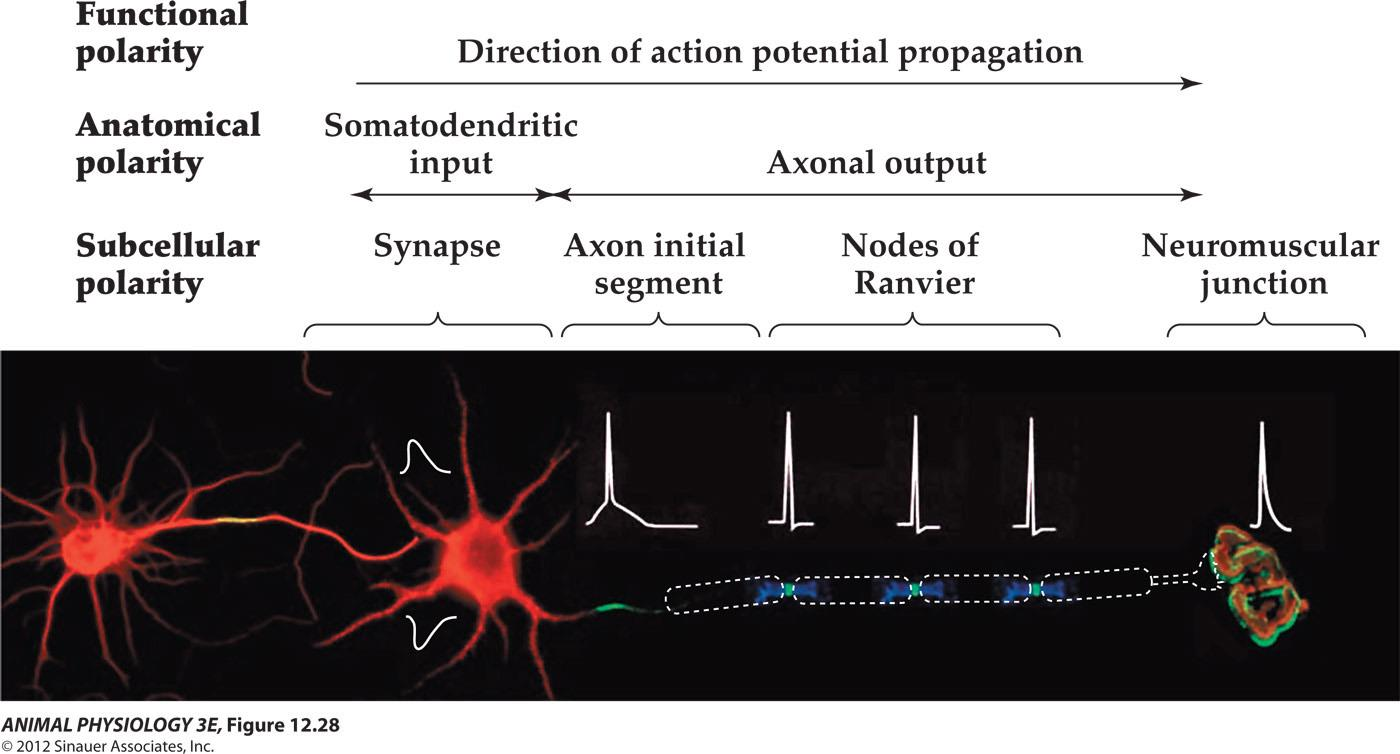
- Na channels (green)
- K channels (blue)
DISCUSSIONS
Suppose you stimulate an axon so that you generate an action potential at both ends at the same instant. Describe the propagation of these action potentials. What happens when they meet?