L4 Circulation - Cardiovascular System
一、The Human Heart
The Basic Structure of Heart
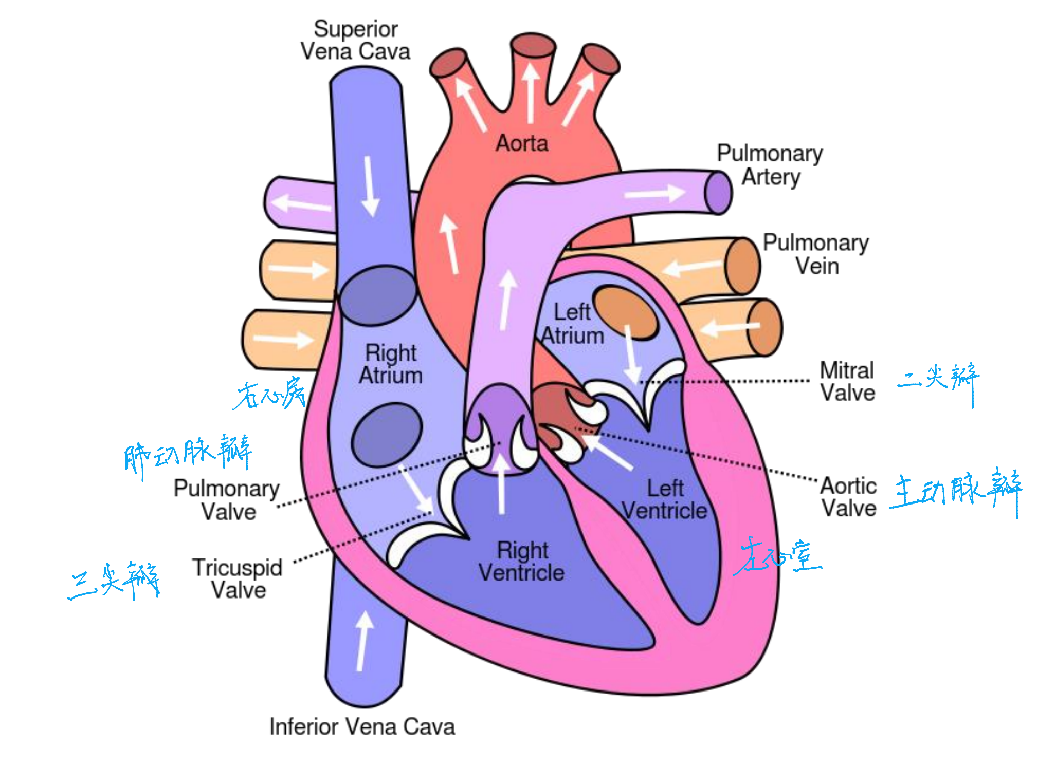
The Blood Circuit
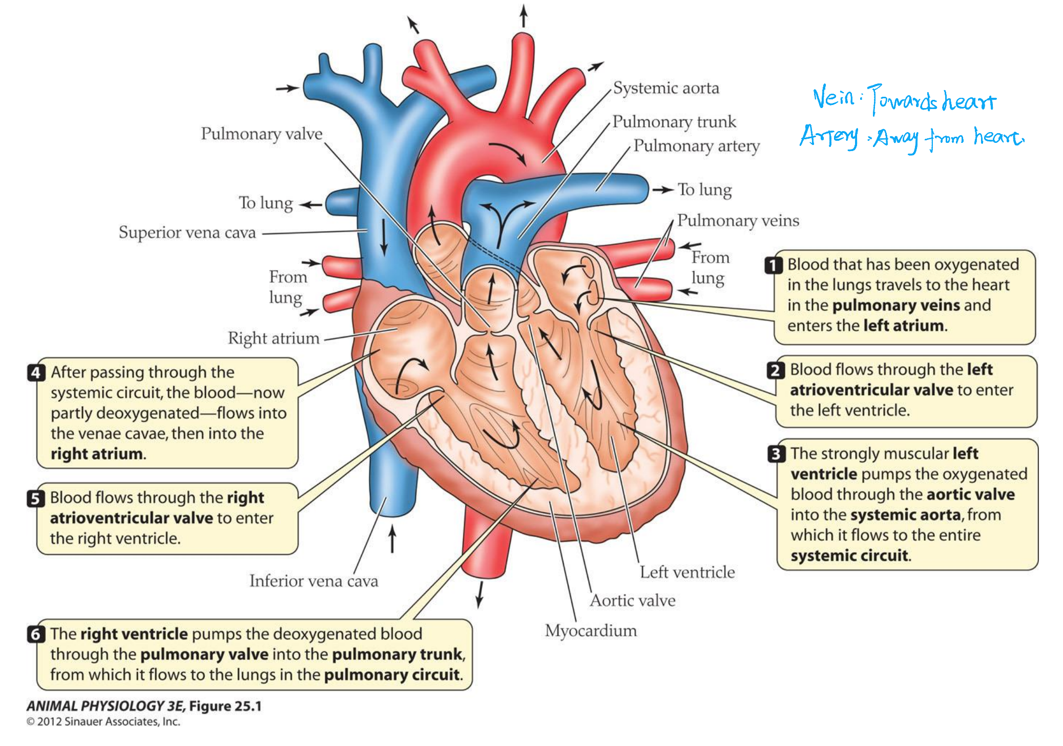
Blood that has been oxygenated in the lungs travels to the heart in the pulmonary veins and enters the left atrium
在肺中被氧化的血液通过肺静脉进入心脏,并进入左心房。
Blood flows through the left atrioventricular valve to enter the left ventricle
血液流经左房室瓣进入左心室.
The strongly muscular left ventricle pumps the oxygenated blood through the aortic valve into the systemic aorta, from which it flows to the entire systemic circuit.
肌肉发达的左心室将含氧血液通过主动脉瓣泵入体循环主动脉,再流向整个体循环。
After passing through the systemic circuit, the blood—now partly deoxygenated—flows into the venae cavae, then into the right atrium
经过全身循环后,血液(现在部分脱氧)流入下腔静脉,然后进入右心房。
Blood flows through the right atrioventricular valve to enter the right ventricle.
血液流经右房室瓣进入右心室。
The right ventricle pumps the deoxygenated blood through the pulmonary valve into the pulmonary trunk from which it flows to the lungs in the pulmonary circuit
右心室将脱氧血液通过肺动脉瓣泵入肺动脉干,再从肺动脉干流入肺循环中的肺。
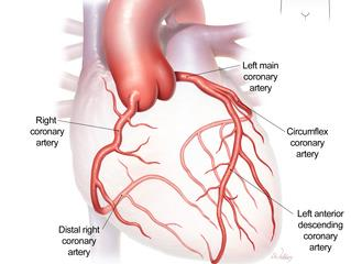
1. The Coronary circulation
The Coronary circulation-delivering O2 to the myocardium
The conducting system and sinoatrial node
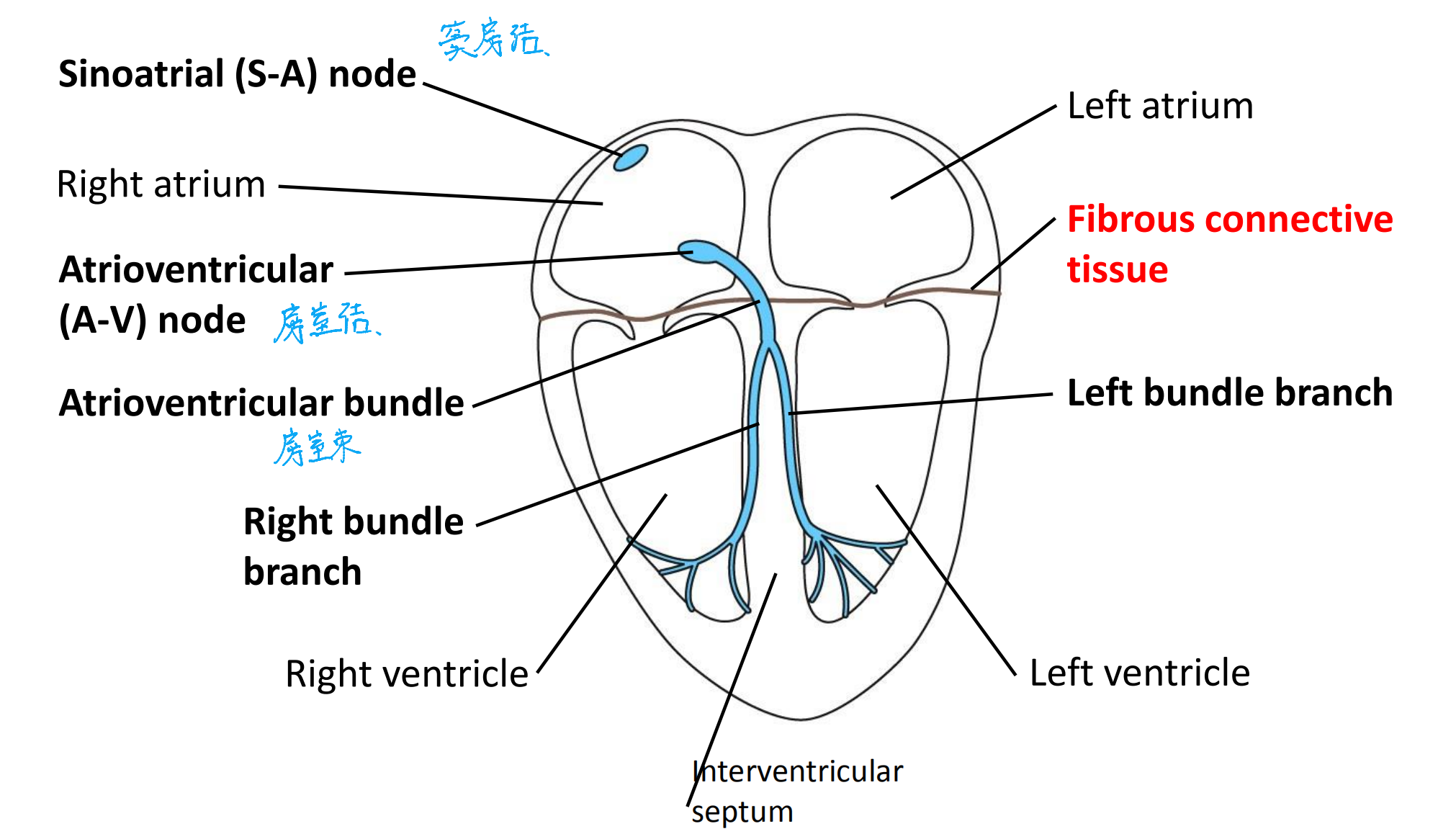
The myocardium of the two atria of the heart is separated from the myocardium of the two ventricles by a layer of fibrous connective tissue across which myocardial cells are not electrically coupled by gap junctions.
The initiation and spread of depolarization during a heartbeat
Conduction: the process by which Depolarization spreads through the Myogeneic heart
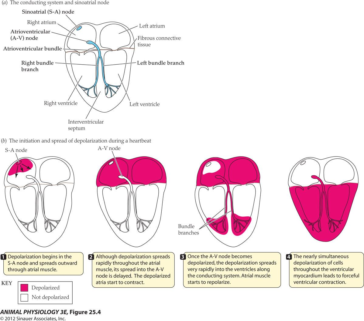
- Depolarization begins in the S-A node and spreads outward through atrial muscle.
- Although depolarization spreads rapidly throughout the atrial muscle, its spread into the A-V node is delayed. The depolarized atria start to contract
- Once the A-V node becomes depolarized, the depolarization spreads very rapidly into the ventricles along the conducting system. Atrial muscle starts to repolarize
- The nearly simultaneous depolarization of cells throughout the ventricular myocardium leads to forceful ventricular contraction
The Dynamics of blood flow, volume, pressure:
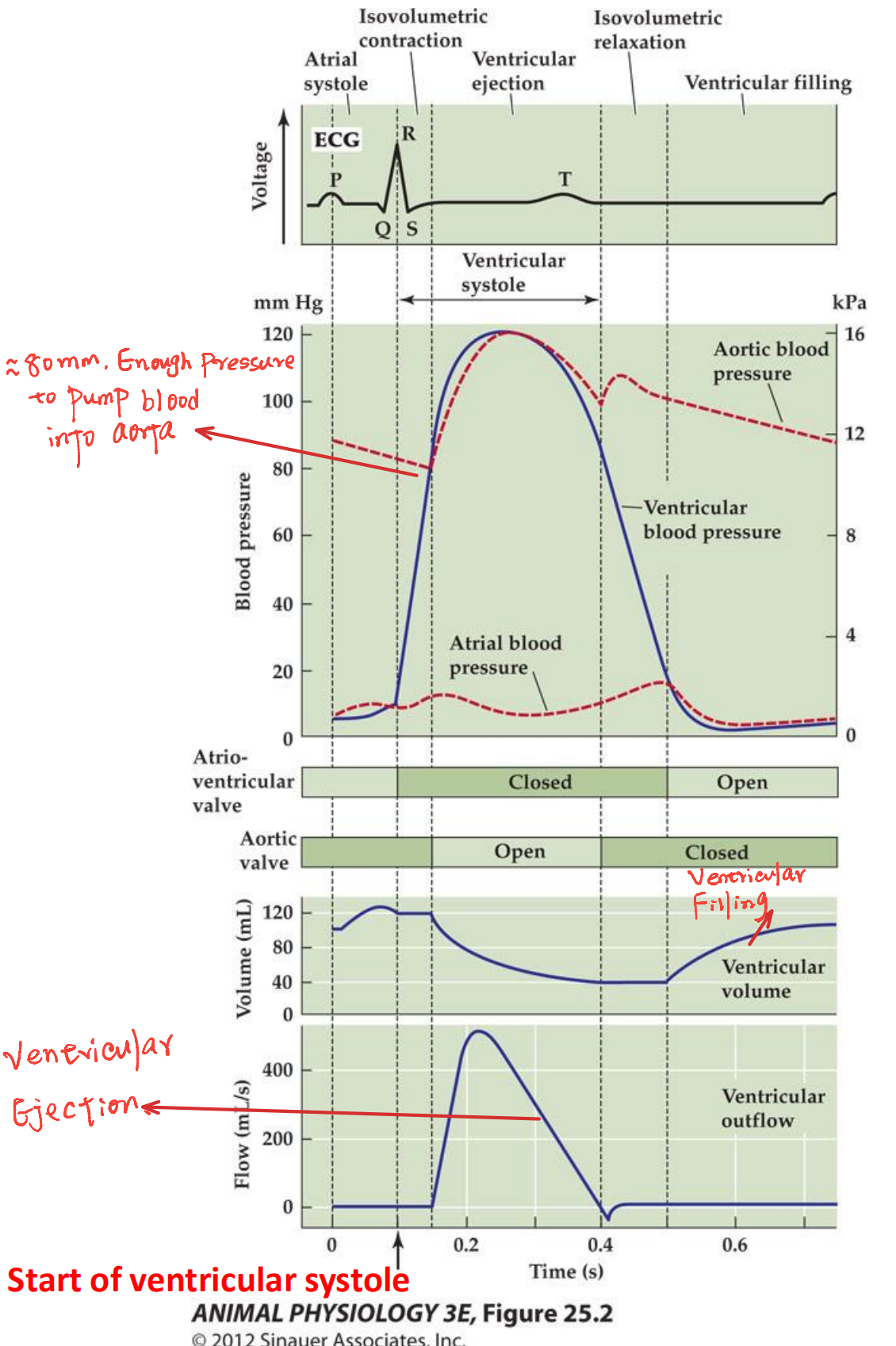
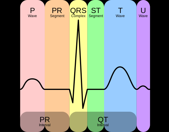
- P: Atrial Depolarization
- P-Q: Atrial Systole
- Q-R-S: Ventrial Depolarization
- R-S: Ventrial Systole
The Heart Cycle:
- Atrial systole 心房收缩
- Isovolumetric contraction 等容收缩
- Ventricular Ejection 心室射血
- Isovolumetric relaxation 等容放松
- Ventricular filling 心室充盈
Cardiac output [(mL/minute)] = heart rate [(beats/minute) ] $\times$ stroke volume [(mL/beat)]
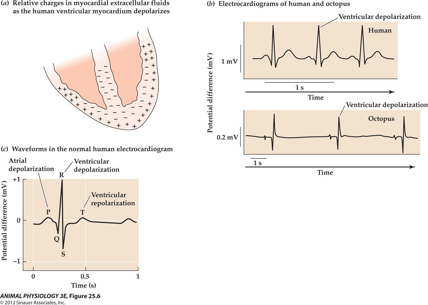
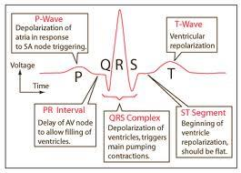
EEG (Electrocardiograms)
Different Systems in Cardiovascular Circulation

Myogenic hearts – vertebrate hearts-they continue to beat without nervous innervations
- Adjacent muscle cells are electrically coupled through gap junctions at intercalating discs
- The pacemaker – a group of specialized muscle cells located on the sinus venous (on the wall of the right atrium) – known as sinoatrial (S-A) node (sinus node).
- S-A node cells – muscle cells, highest frequency of spontaneous depolarization of all cells in the heart, the first to depolarize in the heart. They impose their rhythm of depolarization on the whole heart
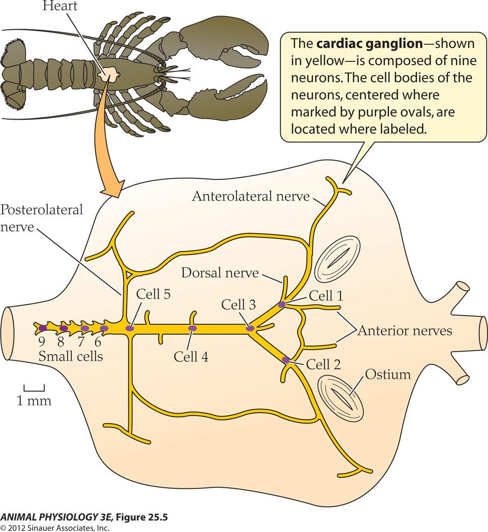
Neurogenic hearts – rhythmic depolarization responsible for initiating the heartbeats originates in nervous tissue (for example, the heart of lobsters).
- 由神经系统控制心脏跳动(例:龙虾)
二、Principals of pressure, resistance and flow in vascular systems
Systole 收缩压 – a period of contraction (pronounced with a long e: sis-tuh-lee)
Diastole 舒张压 – a period of relaxation (dy-as-tuh-lee)
Heart as a pump, we can analyze it in terms of pressure, flow and the volume during these periods.
Blood pressure rises and falls over the heart cycle
- Systolic pressure (resting young adult: 120 mm Hg, 16 Kpa)
- Diastolic pressure (75 mmHg, 10 Pka)
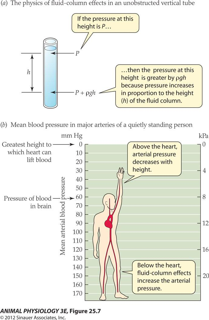
- rho = mess density
- g = acceleration due to gravity
Blood pressure in the arteries of the neck and the head decreases by 10 mm Hg for every 13 cm of height above the heart
Total fluid energy
Total fluid energy: The true driving force for blood flow
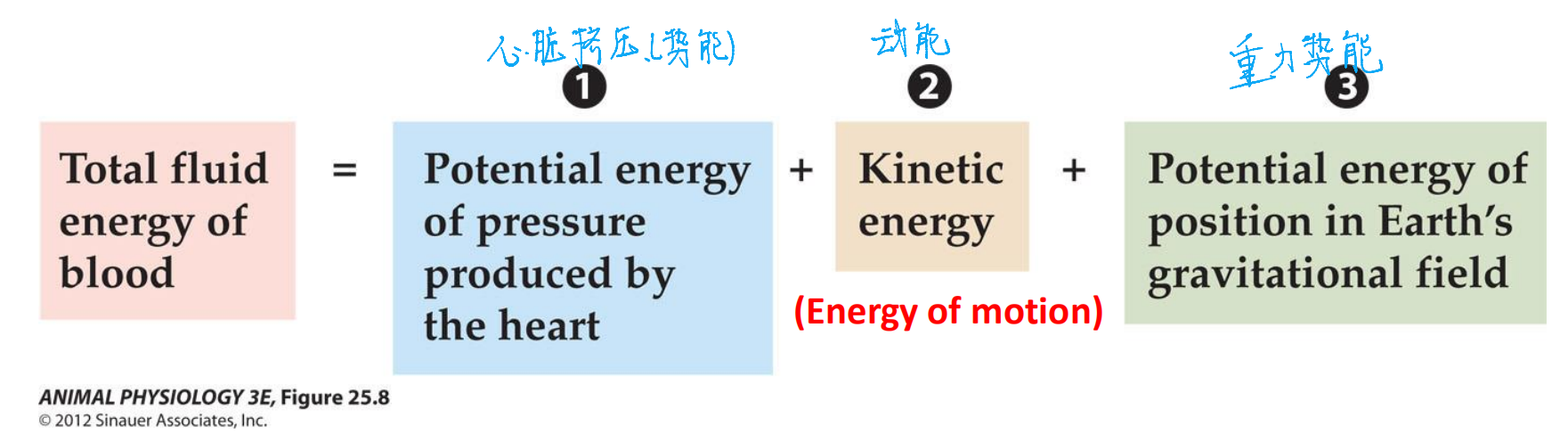
- Blood always flows from where its total fluid energy is higher to where Its total fluid energy is lower.
- Laminar flow (层流) $\rarr$ Velocity (速率)$\rarr$ Viscosity (粘度)
- Internal friction of moving liquid Which degrades kinetic energy into heat Ultimately, pressure is converted to heat
blood pressure and vascular resistance
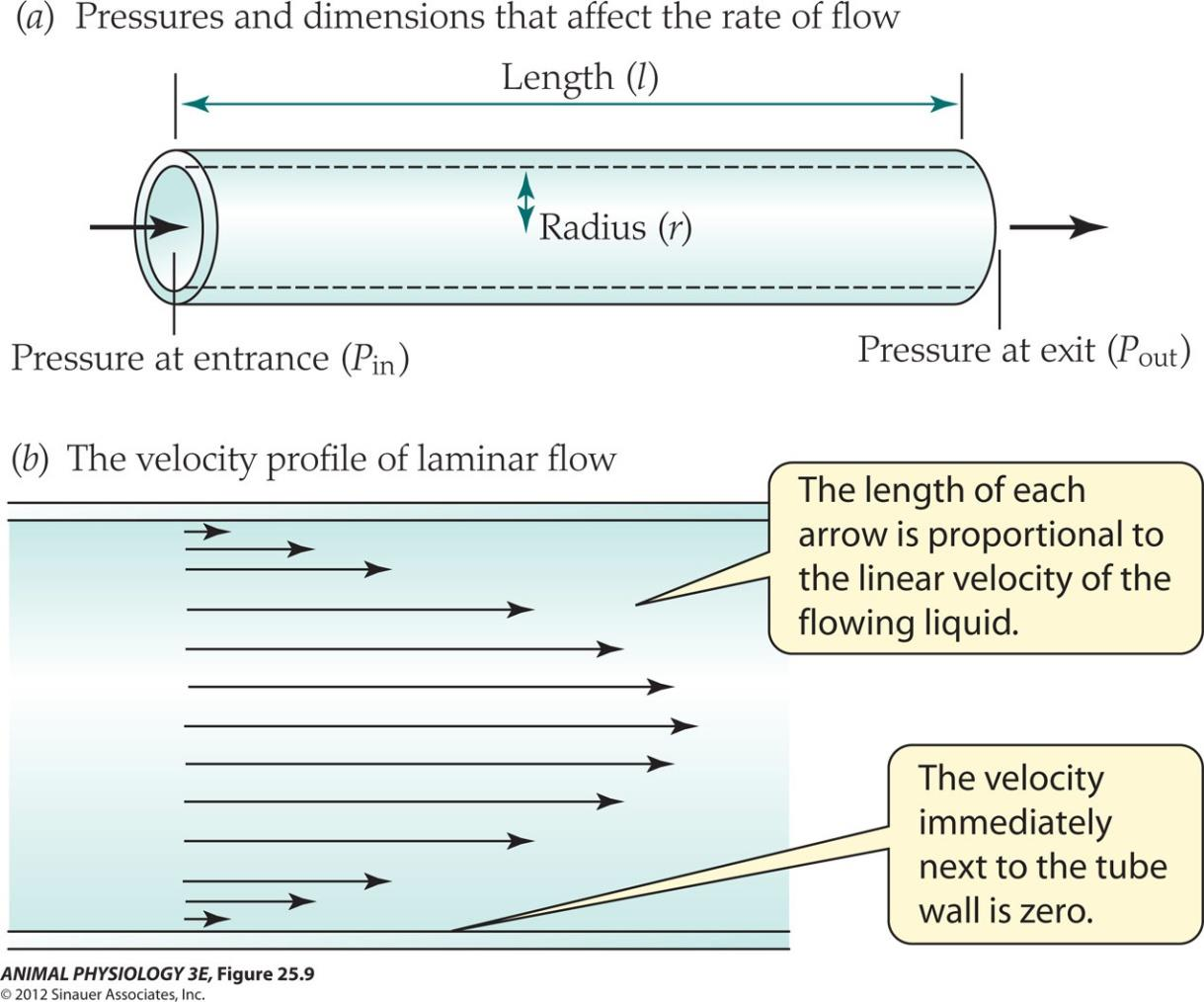
Fluid dynamics, the Hagen-Poiseuille equation is a physical law that gives the pressure drop in a fluid flowing through a long cylindrical pipe
$$
\text{Flow rate} = (P_{in}-P_{out})(π/8)(1/η)(r^{4}
/l) [η = eta]
$$
Another way to describe this is:
$$
\text{Flow rate} = \Delta P/R
$$
(difference in blood pressure; R is the vascular resistance) R = $8η\ l/πr^{4}$
三、Circulation in mammals
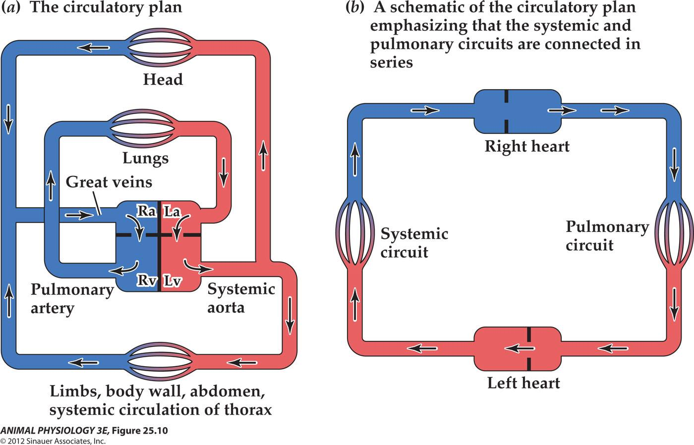
The circulatory plan in mammals and birds
- (a): The circulatory plan as it exists geometrically in the body.
- (b) The same plan, redrawn as a schematic to emphasize the arrangement of the pulmonary and systemic circuits in series with each other. Red and blue portions carry relatively oxygenated and deoxygenated blood, respectively. Ra, right atrium of the heart; La, left atrium; Rv, right ventricle; Lv, left ventricle
The circulatory system
- Closed – blood flows within the discrete vessels
- Open – blood leaves the vessels and bath non-vascular tissue directly
- Both mammals and birds have closed circulatory system
Laplace’s Law
The paradox: the mean blood pressure diminishes hardly at all in the arteries; the arteries become smaller and the walls become thinner
$$
T = r \Delta P
$$
- T= wall tension; r = luminal radius; ∆P = pressure difference across the wall
Even though a small artery may be exposed to the same blood pressure as a large one, the tension developed within its walls is lower than that developed within the walls of the large artery. Accordingly, the walls of small arteries need not be as well fortified as those of large arteries to resist overexpansion. The same principle explains why blood capillaries can be exceedingly thin-walled and yet resist substantial pressures
A microcirculatory bed
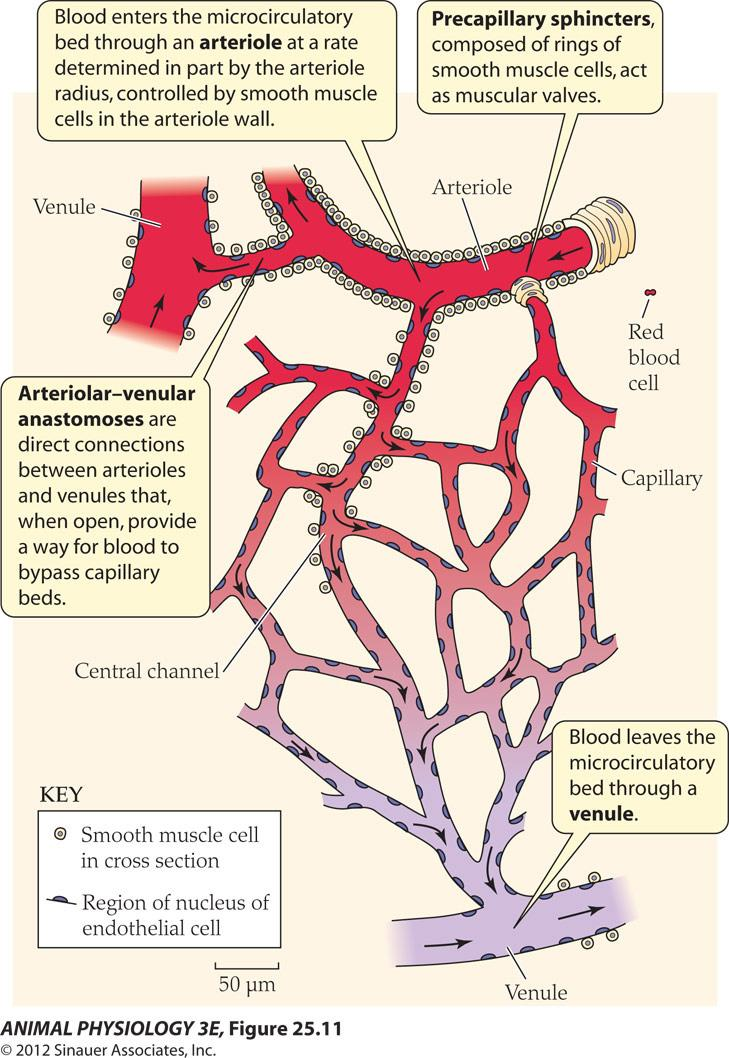
Microcirculatory beds
Three types of vessels:
Arterioles: 30 um diameter, like arteries, vasoconstriction / vasodilation
- (skin changes of environment)
Capillaries: wall has only vascular endothelium Site of O2 exchange, water and other materials Between the blood and the tissues. Capillary bed
Venules: capillary drains into the venules with connective tissues and muscle cells.
Veins
- Thin walled and can not resist high tension
- Contains one-way passive valves
- Skeletal muscles helps to put pressure on veins